 Movie
Movie Controller

[English] 日本語
 Yorodumi
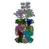
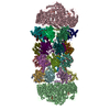
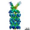
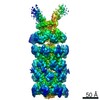
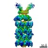
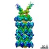
- PDB-7pxc: Substrate-engaged mycobacterial Proteasome-associated ATPase in c... -
+ Open data

- Basic information
| Entry | Database: PDB / ID: 7pxc | ||||||
|---|---|---|---|---|---|---|---|
| Title | Substrate-engaged mycobacterial Proteasome-associated ATPase in complex with open-gate 20S CP - composite map (state A) | ||||||
 Components Components |
| ||||||
 Keywords Keywords | CYTOSOLIC PROTEIN / AAA motor / ATPAse / mycobacterium / proteasome activator / 20S CP | ||||||
| Function / homology |  Function and homology information Function and homology informationubiquitin-like protein reader activity / symbiont defense to host-produced reactive oxygen species / protein pupylation / proteasome-activating nucleotidase complex / response to nitrosative stress / symbiont-mediated perturbation of host defenses / zymogen binding / ATP-dependent peptidase activity / proteasome binding / protein unfolding ...ubiquitin-like protein reader activity / symbiont defense to host-produced reactive oxygen species / protein pupylation / proteasome-activating nucleotidase complex / response to nitrosative stress / symbiont-mediated perturbation of host defenses / zymogen binding / ATP-dependent peptidase activity / proteasome binding / protein unfolding / proteasomal ubiquitin-independent protein catabolic process / proteasome endopeptidase complex / proteasome core complex, beta-subunit complex / threonine-type endopeptidase activity / proteasome core complex, alpha-subunit complex / cellular response to nitric oxide / proteasomal protein catabolic process / peptidoglycan-based cell wall / : / modification-dependent protein catabolic process / protein tag activity / molecular adaptor activity / proteasome-mediated ubiquitin-dependent protein catabolic process / ATP hydrolysis activity / extracellular region / ATP binding / identical protein binding / plasma membrane / cytoplasm Similarity search - Function | ||||||
| Biological species |   Mycobacterium tuberculosis (bacteria) Mycobacterium tuberculosis (bacteria) | ||||||
| Method | ELECTRON MICROSCOPY / single particle reconstruction / cryo EM / Resolution: 3.84 Å | ||||||
 Authors Authors | Jomaa, A. / Kavalchuk, M. / Weber-Ban, E. | ||||||
| Funding support |  Switzerland, 1items Switzerland, 1items
| ||||||
 Citation Citation | Journal: Nat Commun / Year: 2022 Title: Structural basis of prokaryotic ubiquitin-like protein engagement and translocation by the mycobacterial Mpa-proteasome complex. Authors: Mikhail Kavalchuk / Ahmad Jomaa / Andreas U Müller / Eilika Weber-Ban / Abstract: Proteasomes are present in eukaryotes, archaea and Actinobacteria, including the human pathogen Mycobacterium tuberculosis, where proteasomal degradation supports persistence inside the host. In ...Proteasomes are present in eukaryotes, archaea and Actinobacteria, including the human pathogen Mycobacterium tuberculosis, where proteasomal degradation supports persistence inside the host. In mycobacteria and other members of Actinobacteria, prokaryotic ubiquitin-like protein (Pup) serves as a degradation tag post-translationally conjugated to target proteins for their recruitment to the mycobacterial proteasome ATPase (Mpa). Here, we use single-particle cryo-electron microscopy to determine the structure of Mpa in complex with the 20S core particle at an early stage of pupylated substrate recruitment, shedding light on the mechanism of substrate translocation. Two conformational states of Mpa show how substrate is translocated stepwise towards the degradation chamber of the proteasome core particle. We also demonstrate, in vitro and in vivo, the importance of a structural feature in Mpa that allows formation of alternating charge-complementary interactions with the proteasome resulting in radial, rail-guided movements during the ATPase conformational cycle. | ||||||
| History |
|
- Structure visualization
| Movie |
Movie viewer |
|---|---|
| Structure viewer | Molecule: MolmilJmol/JSmol |
- Downloads & links
-Download
| PDBx/mmCIF format | 7pxc.cif.gz | 1.6 MB | Display | PDBx/mmCIF format |
|---|---|---|---|---|
| PDB format | pdb7pxc.ent.gz | Display | PDB format | |
| PDBx/mmJSON format | 7pxc.json.gz | Tree view | PDBx/mmJSON format | |
| Others |  Other downloads Other downloads |
-Validation report
| Arichive directory | https://data.pdbj.org/pub/pdb/validation_reports/px/7pxcftp://data.pdbj.org/pub/pdb/validation_reports/px/7pxc | HTTPS FTP |
|---|
-Related structure data
| Related structure data |  13697MC  7px9C  7pxaC  7pxbC  7pxdC M: map data used to model this data C: citing same article ( |
|---|---|
| Similar structure data |


-Links
 PDBj
PDBj


- Assembly
| Deposited unit | 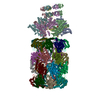
|
|---|---|
| 1 |
|
-Components
-Proteasome subunit ... , 2 types, 28 molecules 02468IKOQTXZdfHJLMNPRSUVWYab
| #1: Protein | Mass: 26911.039 Da / Num. of mol.: 14 Source method: isolated from a genetically manipulated source Details: The first 7 residues were removed (open gate proteasome) Source: (gene. exp.) Mycobacterium tuberculosis (strain ATCC 25618 / H37Rv) (bacteria)Strain: ATCC 25618 / H37Rv / Gene: prcA, Rv2109c / Production host: #4: Protein | Mass: 30332.006 Da / Num. of mol.: 14 Source method: isolated from a genetically manipulated source Source: (gene. exp.) Mycobacterium tuberculosis (strain ATCC 25618 / H37Rv) (bacteria)Strain: ATCC 25618 / H37Rv / Gene: prcB, Rv2110c / Production host: References: UniProt: P9WHT9, proteasome endopeptidase complex |
|---|
-Protein , 2 types, 8 molecules 1ABCDEFG
| #2: Protein | Mass: 67487.930 Da / Num. of mol.: 7 Source method: isolated from a genetically manipulated source Details: Mpa C-terminal extension containing GQYL motif which interacts with the proteasome Source: (gene. exp.) Mycobacterium tuberculosis (strain ATCC 25618 / H37Rv) (bacteria)Strain: ATCC 25618 / H37Rv / Gene: mpa, Rv2115c, MTCY261.11c / Production host: #3: Protein | | Mass: 7095.416 Da / Num. of mol.: 1 Source method: isolated from a genetically manipulated source Source: (gene. exp.) Mycobacterium tuberculosis (strain ATCC 25618 / H37Rv) (bacteria)Strain: ATCC 25618 / H37Rv / Gene: pup, Rv2111c / Production host: |
|---|
-Non-polymers , 3 types, 11 molecules 


| #5: Chemical | ChemComp-ATP /  Mass: 507.181 Da / Num. of mol.: 5 / Source method: obtained synthetically / Formula: C10H16N5O13P3 / Feature type: SUBJECT OF INVESTIGATION / Comment: ATP, energy-carrying molecule*YM Mass: 507.181 Da / Num. of mol.: 5 / Source method: obtained synthetically / Formula: C10H16N5O13P3 / Feature type: SUBJECT OF INVESTIGATION / Comment: ATP, energy-carrying molecule*YM#6: Chemical | ChemComp-MG /  Mass: 24.305 Da / Num. of mol.: 5 / Source method: obtained synthetically / Formula: Mg / Feature type: SUBJECT OF INVESTIGATION Mass: 24.305 Da / Num. of mol.: 5 / Source method: obtained synthetically / Formula: Mg / Feature type: SUBJECT OF INVESTIGATION#7: Chemical | ChemComp-ADP / |  Mass: 427.201 Da / Num. of mol.: 1 / Source method: obtained synthetically / Formula: C10H15N5O10P2 / Feature type: SUBJECT OF INVESTIGATION / Comment: ADP, energy-carrying molecule*YM Mass: 427.201 Da / Num. of mol.: 1 / Source method: obtained synthetically / Formula: C10H15N5O10P2 / Feature type: SUBJECT OF INVESTIGATION / Comment: ADP, energy-carrying molecule*YM |
|---|
-Details
| Has ligand of interest | Y |
|---|
-Experimental details
-Experiment
| Experiment | Method: ELECTRON MICROSCOPY |
|---|---|
| EM experiment | Aggregation state: PARTICLE / 3D reconstruction method: single particle reconstruction |
- Sample preparation
| Component | Name: Mycobacterial Proteasome-associated ATPase in complex with substrate and open-gate 20SCP Type: COMPLEX / Entity ID: #1-#2 / Source: RECOMBINANT |
|---|---|
| Molecular weight | Value: 1.1 MDa / Experimental value: NO |
| Source (natural) | Organism: Mycobacterium tuberculosis (bacteria) |
| Source (recombinant) | Organism: |
| Buffer solution | pH: 7.5 |
| Specimen | Embedding applied: NO / Shadowing applied: NO / Staining applied: NO / Vitrification applied: YES |
| Vitrification | Instrument: FEI VITROBOT MARK IV / Cryogen name: ETHANE-PROPANE |
- Electron microscopy imaging
| Experimental equipment |  Model: Titan Krios / Image courtesy: FEI Company |
|---|---|
| Microscopy | Model: FEI TITAN KRIOS |
| Electron gun | Electron source:  FIELD EMISSION GUN / Accelerating voltage: 300 kV / Illumination mode: FLOOD BEAM FIELD EMISSION GUN / Accelerating voltage: 300 kV / Illumination mode: FLOOD BEAM |
| Electron lens | Mode: BRIGHT FIELD / Nominal magnification: 105000 X / Nominal defocus max: 2500 nm / Nominal defocus min: 1500 nm / Alignment procedure: COMA FREE |
| Specimen holder | Cryogen: NITROGEN / Specimen holder model: FEI TITAN KRIOS AUTOGRID HOLDER |
| Image recording | Electron dose: 50 e/Å2 / Film or detector model: GATAN K3 (6k x 4k) |
- Processing
| EM software |
| |||||||||||||||||||||||||||
|---|---|---|---|---|---|---|---|---|---|---|---|---|---|---|---|---|---|---|---|---|---|---|---|---|---|---|---|---|
| CTF correction | Type: PHASE FLIPPING AND AMPLITUDE CORRECTION | |||||||||||||||||||||||||||
| Particle selection | Num. of particles selected: 860718 | |||||||||||||||||||||||||||
| 3D reconstruction | Resolution: 3.84 Å / Resolution method: OTHER / Num. of particles: 48054 / Symmetry type: POINT | |||||||||||||||||||||||||||
| Atomic model building | Protocol: RIGID BODY FIT / Space: REAL | |||||||||||||||||||||||||||
| Atomic model building | PDB-ID: 5KWA Accession code: 5KWA / Source name: PDB / Type: experimental model |

