 Movie
Movie Controller

[English] 日本語
 Yorodumi
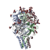
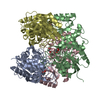
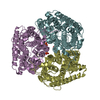
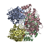
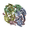
- EMDB-13668: C3-focused map on the ectodomain of the Lassa virus spike complex -
+ Open data

- Basic information
| Entry | Database: EMDB / ID: EMD-13668 | |||||||||
|---|---|---|---|---|---|---|---|---|---|---|
| Title | C3-focused map on the ectodomain of the Lassa virus spike complex | |||||||||
 Map data Map data | C3-symmetric focused map | |||||||||
 Sample Sample |
| |||||||||
| Function / homology |  Function and homology information Function and homology informationhost cell Golgi membrane / receptor-mediated endocytosis of virus by host cell / host cell endoplasmic reticulum membrane / fusion of virus membrane with host endosome membrane / viral envelope / virion attachment to host cell / host cell plasma membrane / virion membrane / membrane / metal ion binding Similarity search - Function | |||||||||
| Biological species |  Lassa virus Josiah Lassa virus Josiah | |||||||||
| Method | single particle reconstruction / cryo EM / Resolution: 2.3 Å | |||||||||
 Authors Authors | Diskin R / Katz M | |||||||||
| Funding support | 1 items
| |||||||||
 Citation Citation | Journal: Nature / Year: 2022 Title: Structure and receptor recognition by the Lassa virus spike complex. Authors: Michael Katz / Jonathan Weinstein / Maayan Eilon-Ashkenazy / Katrin Gehring / Hadas Cohen-Dvashi / Nadav Elad / Sarel J Fleishman / Ron Diskin /  Abstract: Lassa virus (LASV) is a human pathogen, causing substantial morbidity and mortality. Similar to other Arenaviridae, it presents a class-I spike complex on its surface that facilitates cell entry. The ...Lassa virus (LASV) is a human pathogen, causing substantial morbidity and mortality. Similar to other Arenaviridae, it presents a class-I spike complex on its surface that facilitates cell entry. The virus's cellular receptor is matriglycan, a linear carbohydrate that is present on α-dystroglycan, but the molecular mechanism that LASV uses to recognize this glycan is unknown. In addition, LASV and other arenaviruses have a unique signal peptide that forms an integral and functionally important part of the mature spike; yet the structure, function and topology of the signal peptide in the membrane remain uncertain. Here we solve the structure of a complete native LASV spike complex, finding that the signal peptide crosses the membrane once and that its amino terminus is located in the extracellular region. Together with a double-sided domain-switching mechanism, the signal peptide helps to stabilize the spike complex in its native conformation. This structure reveals that the LASV spike complex is preloaded with matriglycan, suggesting the mechanism of binding and rationalizing receptor recognition by α-dystroglycan-tropic arenaviruses. This discovery further informs us about the mechanism of viral egress and may facilitate the rational design of novel therapeutics that exploit this binding site. | |||||||||
| History |
|
- Structure visualization
| Movie |
Movie viewer |
|---|---|
| Structure viewer | EM map: SurfViewMolmilJmol/JSmol |
| Supplemental images |

- Downloads & links
-EMDB archive
| Map data | emd_13668.map.gz | 32.3 MB | EMDB map data format | |
|---|---|---|---|---|
| Header (meta data) | emd-13668-v30.xmlemd-13668.xml | 13.7 KB 13.7 KB | Display Display | EMDB header |
| FSC (resolution estimation) | emd_13668_fsc.xml | 8.9 KB | Display | FSC data file |
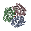
| Images |  emd_13668.png emd_13668.png | 137 KB | ||
| Masks | emd_13668_msk_1.map | 64 MB | Mask map | |
| Others | emd_13668_half_map_1.map.gzemd_13668_half_map_2.map.gz | 59.4 MB 59.4 MB | ||
| Archive directory |  http://ftp.pdbj.org/pub/emdb/structures/EMD-13668ftp://ftp.pdbj.org/pub/emdb/structures/EMD-13668 http://ftp.pdbj.org/pub/emdb/structures/EMD-13668ftp://ftp.pdbj.org/pub/emdb/structures/EMD-13668 | HTTPS FTP |
-Related structure data


-Links
| EMDB pages | EMDB (EBI/PDBe) / EMDataResource |
|---|
-Map
| File | Download / File: emd_13668.map.gz / Format: CCP4 / Size: 64 MB / Type: IMAGE STORED AS FLOATING POINT NUMBER (4 BYTES) | ||||||||||||||||||||||||||||||||||||||||||||||||||||||||||||
|---|---|---|---|---|---|---|---|---|---|---|---|---|---|---|---|---|---|---|---|---|---|---|---|---|---|---|---|---|---|---|---|---|---|---|---|---|---|---|---|---|---|---|---|---|---|---|---|---|---|---|---|---|---|---|---|---|---|---|---|---|---|
| Annotation | C3-symmetric focused map | ||||||||||||||||||||||||||||||||||||||||||||||||||||||||||||
| Projections & slices | Image control
Images are generated by Spider. | ||||||||||||||||||||||||||||||||||||||||||||||||||||||||||||
| Voxel size | X=Y=Z: 1.038 Å | ||||||||||||||||||||||||||||||||||||||||||||||||||||||||||||
| Density |
| ||||||||||||||||||||||||||||||||||||||||||||||||||||||||||||
| Symmetry | Space group: 1 | ||||||||||||||||||||||||||||||||||||||||||||||||||||||||||||
| Details | EMDB XML:
CCP4 map header:
| ||||||||||||||||||||||||||||||||||||||||||||||||||||||||||||
 Z (Sec.)
 Y (Row.)
 X (Col.)


-Supplemental data
-Mask #1
| File | emd_13668_msk_1.map | ||||||||||||
|---|---|---|---|---|---|---|---|---|---|---|---|---|---|
| Projections & Slices |
| ||||||||||||
| Density Histograms |


-Half map: Half map A
| File | emd_13668_half_map_1.map | ||||||||||||
|---|---|---|---|---|---|---|---|---|---|---|---|---|---|
| Annotation | Half map A | ||||||||||||
| Projections & Slices |
| ||||||||||||
| Density Histograms |


-Half map: Half map B
| File | emd_13668_half_map_2.map | ||||||||||||
|---|---|---|---|---|---|---|---|---|---|---|---|---|---|
| Annotation | Half map B | ||||||||||||
| Projections & Slices |
| ||||||||||||
| Density Histograms |


- Sample components
-Entire : The complete spike complex
| Entire | Name: The complete spike complex |
|---|---|
| Components |
|
-Supramolecule #1: The complete spike complex
| Supramolecule | Name: The complete spike complex / type: complex / ID: 1 / Parent: 0 / Macromolecule list: all |
|---|---|
| Source (natural) | Organism: Lassa virus Josiah |
| Recombinant expression | Organism:  Homo sapiens (human) Homo sapiens (human) |
-Macromolecule #1: Membrane-soluble trimeric spike complex from the Lassa virus
| Macromolecule | Name: Membrane-soluble trimeric spike complex from the Lassa virus type: protein_or_peptide / ID: 1 / Enantiomer: LEVO |
|---|---|
| Source (natural) | Organism: Lassa virus Josiah |
| Recombinant expression | Organism: Homo sapiens (human) |
| Sequence | String: MGQIVTFFQE VPHVIEEVMN IVLIALSVLA VLKGLYNFAT CGLVGLVTFL LLCGRSCTTS LYKGVYELQ TLELNMETLN MTMPLSCTKN NSHHYIMVGN ETGLELTLTN TSIINHKFCN L SDAHKKNL YDHALMSIIS TFHLSIPNFN QYEAMSCDFN GGKISVQYNL ...String: MGQIVTFFQE VPHVIEEVMN IVLIALSVLA VLKGLYNFAT CGLVGLVTFL LLCGRSCTTS LYKGVYELQ TLELNMETLN MTMPLSCTKN NSHHYIMVGN ETGLELTLTN TSIINHKFCN L SDAHKKNL YDHALMSIIS TFHLSIPNFN QYEAMSCDFN GGKISVQYNL SHSYAGDAAN HC GTVANGV LQTFMRMAWG GSYIALDSGR GNWDCIMTSY QYLIIQNTTW EDHCQFSRPS PIG YLGLLS QRTRDIYISR RLLGTFTWTL SDSEGKDTPG GYCLTRWMLI EAELKCFGNT AVAK CNEKH DEEFCDMLRL FDFNKQAIQR LKAEAQMSIQ LINKAVNALI NDQLIMKNHL RDIMG IPYC NYSKYWYLNH TTTGRTSLPK CWLVSNGSYL NETHFSDDIE QQADNMITEM LQKEYM ERQ GKTPLGLVDL FVFSTSFYLI SIFLHLVKIP THRHIVGKSC PKPHRLNHMG ICSCGLY KQ PGVPVKWKRG GGSDYKDDDD K |
-Experimental details
-Structure determination
| Method | cryo EM |
|---|---|
 Processing Processing | single particle reconstruction |
| Aggregation state | particle |
-Sample preparation
| Buffer | pH: 7.4 |
|---|---|
| Vitrification | Cryogen name: ETHANE |
- Electron microscopy
| Microscope | FEI TITAN KRIOS |
|---|---|
| Image recording | Film or detector model: GATAN K3 (6k x 4k) / Average electron dose: 73.0 e/Å2 |
| Electron beam | Acceleration voltage: 300 kV / Electron source:  FIELD EMISSION GUN FIELD EMISSION GUN |
| Electron optics | Illumination mode: FLOOD BEAM / Imaging mode: BRIGHT FIELD |
| Experimental equipment |  Model: Titan Krios / Image courtesy: FEI Company |

