 Movie
Movie Controller

[English] 日本語
 Yorodumi
- EMDB-13287: polysome, ribosome pair (conformation 1) in Mycoplasma pneumoniae... -
+ Open data

- Basic information
| Entry |  | |||||||||
|---|---|---|---|---|---|---|---|---|---|---|
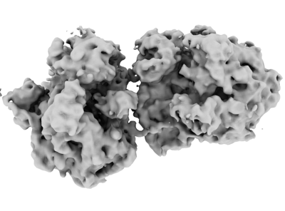
| Title | polysome, ribosome pair (conformation 1) in Mycoplasma pneumoniae cells | |||||||||
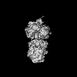
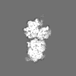
 Map data Map data | ||||||||||
 Sample Sample |
| |||||||||
| Biological species |  Mycoplasma pneumoniae (strain ATCC 29342 / M129) (bacteria) Mycoplasma pneumoniae (strain ATCC 29342 / M129) (bacteria) | |||||||||
| Method | subtomogram averaging / cryo EM / Resolution: 12.0 Å | |||||||||
 Authors Authors | Xue L / Lenz S / Rappsilber J / Mahamid J | |||||||||
| Funding support |  Germany, 1 items Germany, 1 items
| |||||||||
 Citation Citation | Journal: Nature / Year: 2022 Title: Visualizing translation dynamics at atomic detail inside a bacterial cell. Authors: Liang Xue / Swantje Lenz / Maria Zimmermann-Kogadeeva / Dimitry Tegunov / Patrick Cramer / Peer Bork / Juri Rappsilber / Julia Mahamid /   Abstract: Translation is the fundamental process of protein synthesis and is catalysed by the ribosome in all living cells. Here we use advances in cryo-electron tomography and sub-tomogram analysis to ...Translation is the fundamental process of protein synthesis and is catalysed by the ribosome in all living cells. Here we use advances in cryo-electron tomography and sub-tomogram analysis to visualize the structural dynamics of translation inside the bacterium Mycoplasma pneumoniae. To interpret the functional states in detail, we first obtain a high-resolution in-cell average map of all translating ribosomes and build an atomic model for the M. pneumoniae ribosome that reveals distinct extensions of ribosomal proteins. Classification then resolves 13 ribosome states that differ in their conformation and composition. These recapitulate major states that were previously resolved in vitro, and reflect intermediates during active translation. On the basis of these states, we animate translation elongation inside native cells and show how antibiotics reshape the cellular translation landscapes. During translation elongation, ribosomes often assemble in defined three-dimensional arrangements to form polysomes. By mapping the intracellular organization of translating ribosomes, we show that their association into polysomes involves a local coordination mechanism that is mediated by the ribosomal protein L9. We propose that an extended conformation of L9 within polysomes mitigates collisions to facilitate translation fidelity. Our work thus demonstrates the feasibility of visualizing molecular processes at atomic detail inside cells. #1: Journal: Biorxiv / Year: 2021Title: Visualizing translation dynamics at atomic detail inside a bacterial cell Authors: Xue L / Lenz S / Zimmermann-Kogadeeva M / Tegunov D / Cramer P / Bork P / Rappsilber J / Mahamid J | |||||||||
| History |
|
- Structure visualization
| Supplemental images |
|---|
- Downloads & links
-EMDB archive
| Map data | emd_13287.map.gz | 4.8 MB |  EMDB map data format EMDB map data format | |
|---|---|---|---|---|
| Header (meta data) | emd-13287-v30.xmlemd-13287.xml | 22.4 KB 22.4 KB | Display Display | EMDB header |
| FSC (resolution estimation) | emd_13287_fsc.xml | 9.2 KB | Display | FSC data file |
| Images |  emd_13287.png emd_13287.png | 62.9 KB | ||
| Masks | emd_13287_msk_1.map | 64 MB | Mask map | |
| Others | emd_13287_half_map_1.map.gzemd_13287_half_map_2.map.gz | 49.6 MB 49.6 MB | ||
| Archive directory |  http://ftp.pdbj.org/pub/emdb/structures/EMD-13287ftp://ftp.pdbj.org/pub/emdb/structures/EMD-13287 http://ftp.pdbj.org/pub/emdb/structures/EMD-13287ftp://ftp.pdbj.org/pub/emdb/structures/EMD-13287 | HTTPS FTP |
-Validation report
| Summary document | emd_13287_validation.pdf.gz | 801.2 KB | Display | EMDB validaton report |
|---|---|---|---|---|
| Full document | emd_13287_full_validation.pdf.gz | 800.8 KB | Display | |
| Data in XML | emd_13287_validation.xml.gz | 15.7 KB | Display | |
| Data in CIF | emd_13287_validation.cif.gz | 20.7 KB | Display | |
| Arichive directory | https://ftp.pdbj.org/pub/emdb/validation_reports/EMD-13287ftp://ftp.pdbj.org/pub/emdb/validation_reports/EMD-13287 | HTTPS FTP |
-Related structure data
| Related structure data |  7oocC  7oodC  7p6zC  7pahC 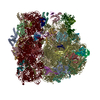 7paiC 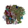 7pajC 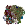 7pakC 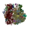 7palC 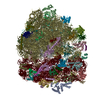 7pamC 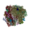 7panC 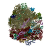 7paoC 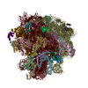 7paqC 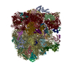 7parC 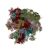 7pasC 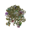 7patC  7pauC 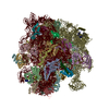 7ph9C 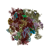 7phaC 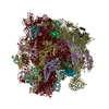 7phbC 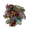 7phcC 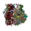 7pi8C  7pi9C  7piaC  7pibC  7picC 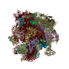 7pioC 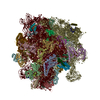 7pipC 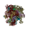 7piqC 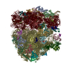 7pirC 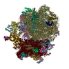 7pisC 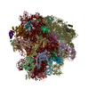 7pitC C: citing same article ( |
|---|


-Links
| EMDB pages | EMDB (EBI/PDBe) / EMDataResource |
|---|
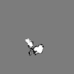
-Map
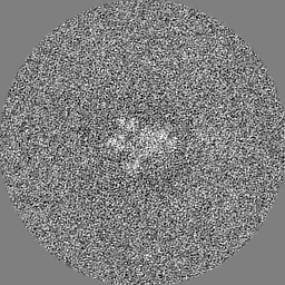
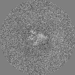
| File | Download / File: emd_13287.map.gz / Format: CCP4 / Size: 64 MB / Type: IMAGE STORED AS FLOATING POINT NUMBER (4 BYTES) | ||||||||||||||||||||||||||||||||||||
|---|---|---|---|---|---|---|---|---|---|---|---|---|---|---|---|---|---|---|---|---|---|---|---|---|---|---|---|---|---|---|---|---|---|---|---|---|---|
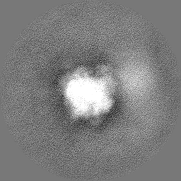
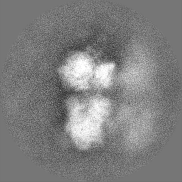
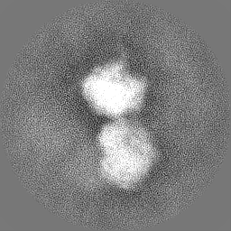
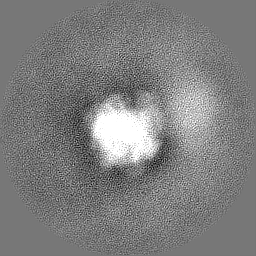
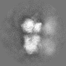
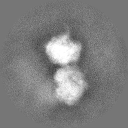
| Projections & slices | Image control
Images are generated by Spider. | ||||||||||||||||||||||||||||||||||||
| Voxel size | X=Y=Z: 3 Å | ||||||||||||||||||||||||||||||||||||
| Density |
| ||||||||||||||||||||||||||||||||||||
| Symmetry | Space group: 1 | ||||||||||||||||||||||||||||||||||||
| Details | EMDB XML:
|
 Z (Sec.)
 Y (Row.)
 X (Col.)


-Supplemental data
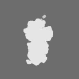
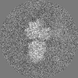
-Mask #1
| File | emd_13287_msk_1.map | ||||||||||||
|---|---|---|---|---|---|---|---|---|---|---|---|---|---|
| Projections & Slices |
| ||||||||||||
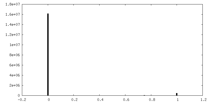
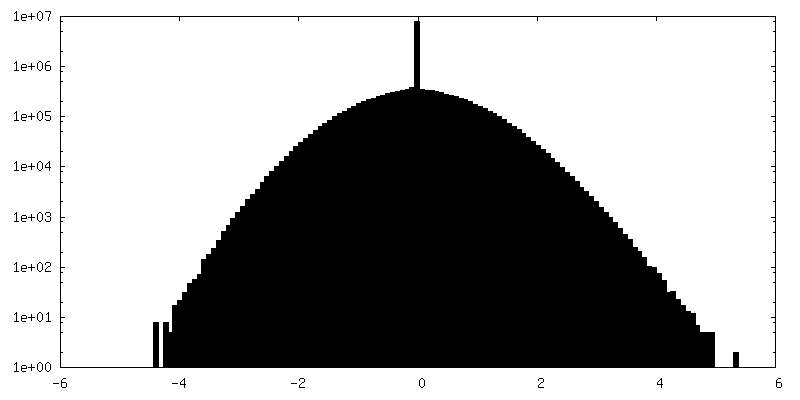
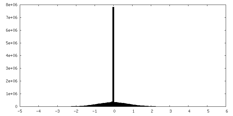
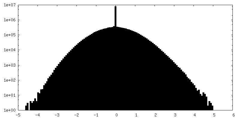
| Density Histograms |

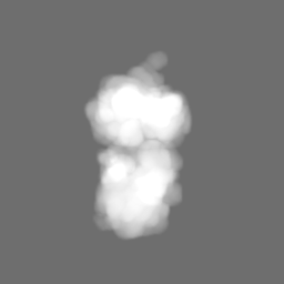


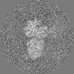
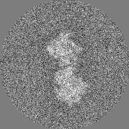
-Half map: #1
| File | emd_13287_half_map_1.map | ||||||||||||
|---|---|---|---|---|---|---|---|---|---|---|---|---|---|
| Projections & Slices |
| ||||||||||||
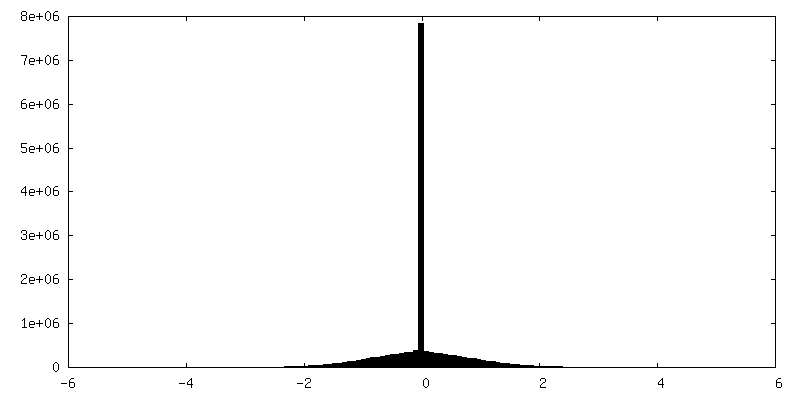
| Density Histograms |
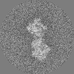
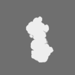
-Half map: #2
| File | emd_13287_half_map_2.map | ||||||||||||
|---|---|---|---|---|---|---|---|---|---|---|---|---|---|
| Projections & Slices |
| ||||||||||||
| Density Histograms |
- Sample components
-Entire : cryo-electron tomograms of untreated Mycoplasma pneumoniae cells
| Entire | Name: cryo-electron tomograms of untreated Mycoplasma pneumoniae cells |
|---|---|
| Components |
|
-Supramolecule #1: cryo-electron tomograms of untreated Mycoplasma pneumoniae cells
| Supramolecule | Name: cryo-electron tomograms of untreated Mycoplasma pneumoniae cells type: organelle_or_cellular_component / ID: 1 / Parent: 0 / Macromolecule list: #1-#55 Details: The tomograms were collected with intact Mycoplasma pneumoniae cells. The sub-tomograms extracted in silico from cellular tomograms are with large box size to accommodate two adjacent ...Details: The tomograms were collected with intact Mycoplasma pneumoniae cells. The sub-tomograms extracted in silico from cellular tomograms are with large box size to accommodate two adjacent ribosomes (one ribosome pair) with polysomes. |
|---|---|
| Source (natural) | Organism: Mycoplasma pneumoniae (strain ATCC 29342 / M129) (bacteria) |
-Experimental details
-Structure determination
| Method | cryo EM |
|---|---|
 Processing Processing | subtomogram averaging |
| Aggregation state | cell |
-Sample preparation
| Buffer | pH: 7.4 |
|---|---|
| Vitrification | Cryogen name: ETHANE-PROPANE / Instrument: HOMEMADE PLUNGER Details: back-side blotting for 2-3 second before plunging using a manual plunger without an environmental chamber. |
| Details | Mycoplasma pneumoniae M129 cells grown on gold Quantifoil grids at 37 degrees Celsius before plunge freezing. |
- Electron microscopy
| Microscope | FEI TITAN KRIOS |
|---|---|
| Image recording | Film or detector model: GATAN K2 SUMMIT (4k x 4k) / Detector mode: COUNTING / Average electron dose: 3.2 e/Å2 |
| Electron beam | Acceleration voltage: 300 kV / Electron source:  FIELD EMISSION GUN FIELD EMISSION GUN |
| Electron optics | Illumination mode: FLOOD BEAM / Imaging mode: BRIGHT FIELD / Cs: 2.7 mm / Nominal defocus max: 3.75 µm / Nominal defocus min: 1.5 µm / Nominal magnification: 81000 |
| Sample stage | Specimen holder model: FEI TITAN KRIOS AUTOGRID HOLDER / Cooling holder cryogen: NITROGEN |
| Experimental equipment |  Model: Titan Krios / Image courtesy: FEI Company |
-Image processing
| Final reconstruction | Applied symmetry - Point group: C1 (asymmetric) / Resolution.type: BY AUTHOR / Resolution: 12.0 Å / Resolution method: FSC 0.143 CUT-OFF / Software - Name: RELION (ver. 3.0.8) / Number subtomograms used: 1154 |
|---|---|
| Extraction | Number tomograms: 356 / Number images used: 77539 Details: Initially 77539 sub-tomograms were extracted from 356 cells, with box size large enough to accommodate a di-ribosome. Then the polysome class with 9616 sub-tomograms were re-centered and re-extracted. |
| Final angle assignment | Type: OTHER |
| FSC plot (resolution estimation) |  |
-Atomic model buiding 1
| Refinement | Space: REAL / Protocol: RIGID BODY FIT |
|---|
