 ムービー
ムービー コントローラー


+ データを開く

- 基本情報
基本情報
| 登録情報 | データベース: EMDB / ID: EMD-1301 | |||||||||
|---|---|---|---|---|---|---|---|---|---|---|
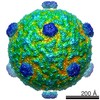
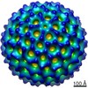
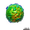
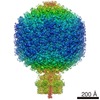
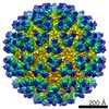
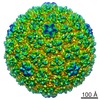
| タイトル | Electron cryomicroscopy comparison of the architectures of the enveloped bacteriophages phi6 and phi8. | |||||||||
 マップデータ マップデータ | 3D reconstruction of the bacteriophage Phi6 virion. | |||||||||
 試料 試料 |
| |||||||||
| 生物種 |  Pseudomonas phage phi6 (ファージ) Pseudomonas phage phi6 (ファージ) | |||||||||
| 手法 | 単粒子再構成法 / クライオ電子顕微鏡法 / 解像度: 18.0 Å | |||||||||
 データ登録者 データ登録者 | Jaalinoja HT / Huiskonen JT / Butcher SJ | |||||||||
 引用 引用 | ジャーナル: Structure / 年: 2007 タイトル: Electron cryomicroscopy comparison of the architectures of the enveloped bacteriophages phi6 and phi8. 著者: Harri T Jäälinoja / Juha T Huiskonen / Sarah J Butcher /  要旨: The enveloped dsRNA bacteriophages phi6 and phi8 are the two most distantly related members of the Cystoviridae family. Their structure and function are similar to that of the Reoviridae but their ...The enveloped dsRNA bacteriophages phi6 and phi8 are the two most distantly related members of the Cystoviridae family. Their structure and function are similar to that of the Reoviridae but their assembly can be conveniently studied in vitro. Electron cryomicroscopy and three-dimensional icosahedral reconstruction were used to determine the structures of the phi6 virion (14 A resolution), phi8 virion (18 A resolution), and phi8 core (8.5 A resolution). Spikes protrude 2 nm from the membrane bilayer in phi6 and 7 nm in phi8. In the phi6 nucleocapsid, 600 copies of P8 and 72 copies of P4 interact with the membrane, whereas in phi8 it is only P4 and 60 copies of a minor protein. The major polymerase complex protein P1 forms a dodecahedral shell from 60 asymmetric dimers in both viruses, but the alpha-helical fold has apparently diverged. These structural differences reflect the different host ranges and entry and assembly mechanisms of the two viruses. | |||||||||
| 履歴 |
|
- 構造の表示
構造の表示
| ムービー |
 ムービービューア ムービービューア |
|---|---|
| 構造ビューア | EMマップ: SurfViewMolmilJmol/JSmol |
| 添付画像 |

- ダウンロードとリンク
-EMDBアーカイブ
| マップデータ | emd_1301.map.gz | 55.5 MB | EMDBマップデータ形式 | |
|---|---|---|---|---|
| ヘッダ (付随情報) | emd-1301-v30.xmlemd-1301.xml | 9.3 KB 9.3 KB | 表示 表示 | EMDBヘッダ |
| 画像 |  1301.gif 1301.gif | 22 KB | ||
| アーカイブディレクトリ |  http://ftp.pdbj.org/pub/emdb/structures/EMD-1301ftp://ftp.pdbj.org/pub/emdb/structures/EMD-1301 http://ftp.pdbj.org/pub/emdb/structures/EMD-1301ftp://ftp.pdbj.org/pub/emdb/structures/EMD-1301 | HTTPS FTP |
-検証レポート
| 文書・要旨 | emd_1301_validation.pdf.gz | 288.2 KB | 表示 | EMDB検証レポート |
|---|---|---|---|---|
| 文書・詳細版 | emd_1301_full_validation.pdf.gz | 287.4 KB | 表示 | |
| XML形式データ | emd_1301_validation.xml.gz | 7.2 KB | 表示 | |
| アーカイブディレクトリ | https://ftp.pdbj.org/pub/emdb/validation_reports/EMD-1301ftp://ftp.pdbj.org/pub/emdb/validation_reports/EMD-1301 | HTTPS FTP |
-関連構造データ

-リンク
| EMDBのページ | EMDB (EBI/PDBe) / EMDataResource |
|---|
-マップ
| ファイル | ダウンロード / ファイル: emd_1301.map.gz / 形式: CCP4 / 大きさ: 127.9 MB / タイプ: IMAGE STORED AS FLOATING POINT NUMBER (4 BYTES) | ||||||||||||||||||||||||||||||||||||||||||||||||||||||||||||||||||||
|---|---|---|---|---|---|---|---|---|---|---|---|---|---|---|---|---|---|---|---|---|---|---|---|---|---|---|---|---|---|---|---|---|---|---|---|---|---|---|---|---|---|---|---|---|---|---|---|---|---|---|---|---|---|---|---|---|---|---|---|---|---|---|---|---|---|---|---|---|---|
| 注釈 | 3D reconstruction of the bacteriophage Phi6 virion. | ||||||||||||||||||||||||||||||||||||||||||||||||||||||||||||||||||||
| 投影像・断面図 | 画像のコントロール
画像は Spider により作成 | ||||||||||||||||||||||||||||||||||||||||||||||||||||||||||||||||||||
| ボクセルのサイズ | X=Y=Z: 2.8 Å | ||||||||||||||||||||||||||||||||||||||||||||||||||||||||||||||||||||
| 密度 |
| ||||||||||||||||||||||||||||||||||||||||||||||||||||||||||||||||||||
| 対称性 | 空間群: 1 | ||||||||||||||||||||||||||||||||||||||||||||||||||||||||||||||||||||
| 詳細 | EMDB XML:
CCP4マップ ヘッダ情報:
| ||||||||||||||||||||||||||||||||||||||||||||||||||||||||||||||||||||
 Z (Sec.)
 Y (Row.)
 X (Col.)


-添付データ
- 試料の構成要素
-全体 : Bacteriophage Phi6 virion
| 全体 | 名称: Bacteriophage Phi6 virion |
|---|---|
| 要素 |
|
-超分子 #1000: Bacteriophage Phi6 virion
| 超分子 | 名称: Bacteriophage Phi6 virion / タイプ: sample / ID: 1000 / 集合状態: complete virion / Number unique components: 1 |
|---|
-超分子 #1: Pseudomonas phage phi6
| 超分子 | 名称: Pseudomonas phage phi6 / タイプ: virus / ID: 1 / Name.synonym: Cystovirus Phi6 virion / NCBI-ID: 10879 / 生物種: Pseudomonas phage phi6 / ウイルスタイプ: VIRION / ウイルス・単離状態: SPECIES / ウイルス・エンベロープ: Yes / ウイルス・中空状態: No / Syn species name: Cystovirus Phi6 virion |
|---|---|
| 宿主 | 生物種: Pseudomonads syringae / 別称: BACTERIA(EUBACTERIA) |
| ウイルス殻 | Shell ID: 1 / 名称: Nucleocapsid / 直径: 550 Å / T番号(三角分割数): 13 |
-実験情報
-構造解析
| 手法 | クライオ電子顕微鏡法 |
|---|---|
 解析 解析 | 単粒子再構成法 |
| 試料の集合状態 | particle |
-試料調製
| 緩衝液 | 詳細: 10 mM potassium phosphate pH 7.5, 1 mM MgCl2 |
|---|---|
| グリッド | 詳細: 400 mesh copper grid, Quantifoil R2/2 holey |
| 凍結 | 凍結剤: ETHANE / チャンバー内温度: 90 K / 装置: HOMEMADE PLUNGER / 詳細: Vitrification instrument: EMBL design 手法: A small vial of ethane is placed inside a larger liquid nitrogen reservoir. The grid holding 3 microliters of the sample is held in place at the bottom of a plunger by the means of fine ...手法: A small vial of ethane is placed inside a larger liquid nitrogen reservoir. The grid holding 3 microliters of the sample is held in place at the bottom of a plunger by the means of fine tweezers. When the liquid ethane is ready, a piece of filter paper is then pressed against the sample to blot off excess buffer, sufficient to leave a thin layer on the grid. The filter paper is removed, and the plunger is allowed to drop into the liquid ethane. Once the grid enters the liquid ethane, the sample is rapidly frozen, and the grid is transferred under liquid nitrogen to a storage box immersed in liquid nitrogen for later use in the microscope. |
- 電子顕微鏡法
| 顕微鏡 | FEI TECNAI F20 |
|---|---|
| 温度 | 最低: 90 K / 最高: 94 K / 平均: 93 K |
| 撮影 | カテゴリ: FILM / フィルム・検出器のモデル: KODAK SO-163 FILM / デジタル化 - スキャナー: ZEISS SCAI / デジタル化 - サンプリング間隔: 7 µm / 実像数: 23 / 詳細: The data was processed at 2.8 ??/pixel sampling. / ビット/ピクセル: 12 |
| Tilt angle min | 0 |
| Tilt angle max | 0 |
| 電子線 | 加速電圧: 200 kV / 電子線源:  FIELD EMISSION GUN FIELD EMISSION GUN |
| 電子光学系 | 倍率(補正後): 49300 / 照射モード: FLOOD BEAM / 撮影モード: BRIGHT FIELD / Cs: 2.0 mm / 最大 デフォーカス(公称値): 2.9 µm / 最小 デフォーカス(公称値): 1.3 µm / 倍率(公称値): 50000 |
| 試料ステージ | 試料ホルダー: Side entry liquid nitrogen-cooled cryo specimen holder 試料ホルダーモデル: GATAN LIQUID NITROGEN |
| 実験機器 |  モデル: Tecnai F20 / 画像提供: FEI Company |
-画像解析
| CTF補正 | 詳細: Each particle, wiener factor 0.1 |
|---|---|
| 最終 再構成 | 想定した対称性 - 点群: I (正20面体型対称) / アルゴリズム: OTHER / 解像度のタイプ: BY AUTHOR / 解像度: 18.0 Å / 解像度の算出法: FSC 0.5 CUT-OFF / ソフトウェア - 名称: pft2, em3dr2, POR, P3DR / 使用した粒子像数: 517 |
