 Movie
Movie Controller

[English] 日本語
 Yorodumi
- EMDB-8005: Resolution and Probabilistic Structural Models of Subcomponents D... -
+ Open data

- Basic information
| Entry | Database: EMDB / ID: EMD-8005 | ||||||||||||
|---|---|---|---|---|---|---|---|---|---|---|---|---|---|
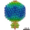
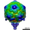
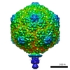
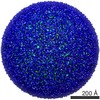
| Title | Resolution and Probabilistic Structural Models of Subcomponents Derived from CryoEM Maps of Mature P22 Bacteriophage | ||||||||||||
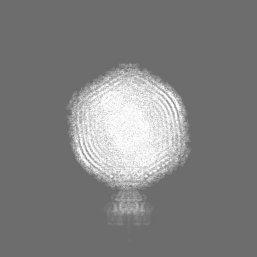
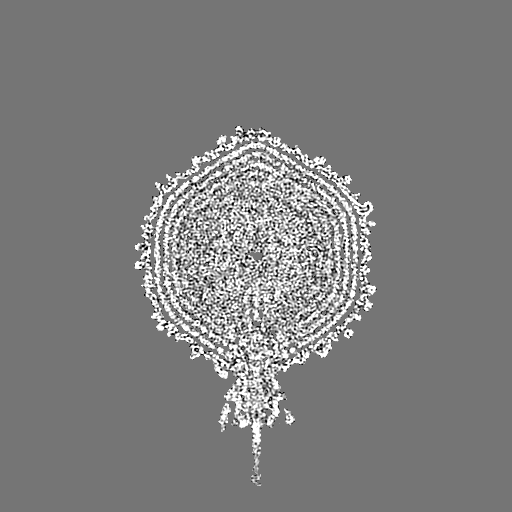
 Map data Map data | P22 mature virion | ||||||||||||
 Sample Sample |
| ||||||||||||
 Keywords Keywords | virion / portal / tailspike / adhesin / VIRAL PROTEIN | ||||||||||||
| Function / homology |  Function and homology information Function and homology informationviral DNA genome packaging, headful / symbiont entry into host cell via disruption of host cell wall peptidoglycan / endo-1,3-alpha-L-rhamnosidase activity / symbiont entry into host cell via disruption of host cell envelope lipopolysaccharide / viral portal complex / symbiont genome ejection through host cell envelope, short tail mechanism / viral DNA genome packaging / virus tail, fiber / symbiont entry into host cell via disruption of host cell envelope / virus tail ...viral DNA genome packaging, headful / symbiont entry into host cell via disruption of host cell wall peptidoglycan / endo-1,3-alpha-L-rhamnosidase activity / symbiont entry into host cell via disruption of host cell envelope lipopolysaccharide / viral portal complex / symbiont genome ejection through host cell envelope, short tail mechanism / viral DNA genome packaging / virus tail, fiber / symbiont entry into host cell via disruption of host cell envelope / virus tail / symbiont entry into host / Hydrolases; Glycosylases; Glycosidases, i.e. enzymes that hydrolyse O- and S-glycosyl compounds / adhesion receptor-mediated virion attachment to host cell / virion assembly / lyase activity / hydrolase activity / virion attachment to host cell Similarity search - Function | ||||||||||||
| Biological species |  Enterobacteria phage P22 (virus) Enterobacteria phage P22 (virus) | ||||||||||||
| Method | single particle reconstruction / cryo EM / Resolution: 10.5 Å | ||||||||||||
 Authors Authors | Pintilie G / Chen DH | ||||||||||||
| Funding support |  United States, 3 items United States, 3 items
| ||||||||||||
 Citation Citation | Journal: Biophys J / Year: 2016 Title: Resolution and Probabilistic Models of Components in CryoEM Maps of Mature P22 Bacteriophage. Authors: Grigore Pintilie / Dong-Hua Chen / Cameron A Haase-Pettingell / Jonathan A King / Wah Chiu / Abstract: CryoEM continues to produce density maps of larger and more complex assemblies with multiple protein components of mixed symmetries. Resolution is not always uniform throughout a cryoEM map, and it ...CryoEM continues to produce density maps of larger and more complex assemblies with multiple protein components of mixed symmetries. Resolution is not always uniform throughout a cryoEM map, and it can be useful to estimate the resolution in specific molecular components of a large assembly. In this study, we present procedures to 1) estimate the resolution in subcomponents by gold-standard Fourier shell correlation (FSC); 2) validate modeling procedures, particularly at medium resolutions, which can include loop modeling and flexible fitting; and 3) build probabilistic models that combine high-accuracy priors (such as crystallographic structures) with medium-resolution cryoEM densities. As an example, we apply these methods to new cryoEM maps of the mature bacteriophage P22, reconstructed without imposing icosahedral symmetry. Resolution estimates based on gold-standard FSC show the highest resolution in the coat region (7.6 Å), whereas other components are at slightly lower resolutions: portal (9.2 Å), hub (8.5 Å), tailspike (10.9 Å), and needle (10.5 Å). These differences are indicative of inherent structural heterogeneity and/or reconstruction accuracy in different subcomponents of the map. Probabilistic models for these subcomponents provide new insights, to our knowledge, and structural information when taking into account uncertainty given the limitations of the observed density. | ||||||||||||
| History |
|
- Structure visualization
| Movie |
Movie viewer |
|---|---|
| Structure viewer | EM map: SurfViewMolmilJmol/JSmol |
| Supplemental images |

- Downloads & links
-EMDB archive
| Map data | emd_8005.map.gz | 24.5 MB | EMDB map data format | |
|---|---|---|---|---|
| Header (meta data) | emd-8005-v30.xmlemd-8005.xml | 37.7 KB 37.7 KB | Display Display | EMDB header |
| FSC (resolution estimation) | emd_8005_fsc_1.xmlemd_8005_fsc_2.xmlemd_8005_fsc_3.xmlemd_8005_fsc_4.xmlemd_8005_fsc_5.xmlemd_8005_fsc_6.xml | 21.2 KB 5.9 KB 21.1 KB 21.3 KB 21.1 KB 21.3 KB | Display Display Display Display Display Display | FSC data file |
| Images |  emd_8005.png emd_8005.png | 161.5 KB | ||
| Filedesc metadata | emd-8005.cif.gz | 7.8 KB | ||
| Others | emd_8005_additional_1.map.gzemd_8005_additional_2.map.gzemd_8005_additional_3.map.gzemd_8005_additional_4.map.gzemd_8005_additional_5.map.gzemd_8005_additional_6.map.gzemd_8005_additional_7.map.gzemd_8005_additional_8.map.gzemd_8005_half_map_1.map.gzemd_8005_half_map_2.map.gz | 465.6 KB 1.2 MB 226.3 KB 24.1 MB 167.4 KB 502.8 KB 582.1 KB 1.4 MB 250.1 MB 250.1 MB | ||
| Archive directory |  http://ftp.pdbj.org/pub/emdb/structures/EMD-8005ftp://ftp.pdbj.org/pub/emdb/structures/EMD-8005 http://ftp.pdbj.org/pub/emdb/structures/EMD-8005ftp://ftp.pdbj.org/pub/emdb/structures/EMD-8005 | HTTPS FTP |
-Related structure data
| Related structure data |  5gaiMC M: atomic model generated by this map C: citing same article ( |
|---|---|
| Similar structure data |


-Links
| EMDB pages | EMDB (EBI/PDBe) / EMDataResource |
|---|
-Map
| File | Download / File: emd_8005.map.gz / Format: CCP4 / Size: 512 MB / Type: IMAGE STORED AS FLOATING POINT NUMBER (4 BYTES) | ||||||||||||||||||||||||||||||||||||||||||||||||||||||||||||||||||||
|---|---|---|---|---|---|---|---|---|---|---|---|---|---|---|---|---|---|---|---|---|---|---|---|---|---|---|---|---|---|---|---|---|---|---|---|---|---|---|---|---|---|---|---|---|---|---|---|---|---|---|---|---|---|---|---|---|---|---|---|---|---|---|---|---|---|---|---|---|---|
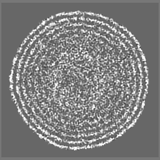
| Annotation | P22 mature virion | ||||||||||||||||||||||||||||||||||||||||||||||||||||||||||||||||||||
| Projections & slices | Image control
Images are generated by Spider. | ||||||||||||||||||||||||||||||||||||||||||||||||||||||||||||||||||||
| Voxel size | X=Y=Z: 2.55 Å | ||||||||||||||||||||||||||||||||||||||||||||||||||||||||||||||||||||
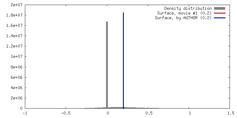
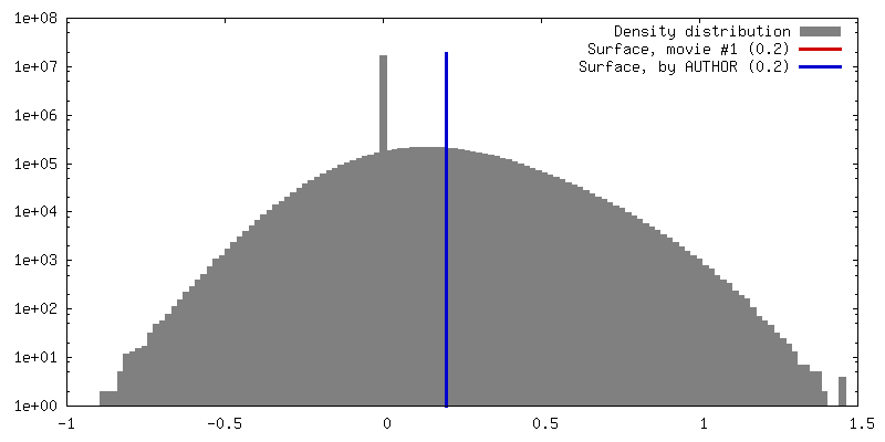
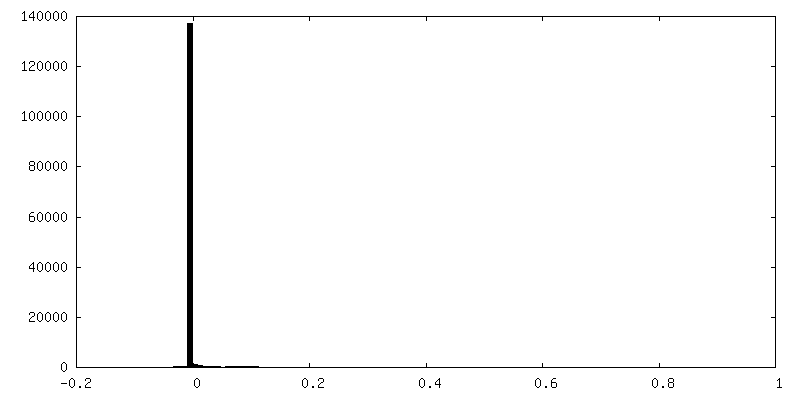
| Density |
| ||||||||||||||||||||||||||||||||||||||||||||||||||||||||||||||||||||
| Symmetry | Space group: 1 | ||||||||||||||||||||||||||||||||||||||||||||||||||||||||||||||||||||
| Details | EMDB XML:
CCP4 map header:
| ||||||||||||||||||||||||||||||||||||||||||||||||||||||||||||||||||||
 Z (Sec.)
 Y (Row.)
 X (Col.)


-Supplemental data
+Additional map: Hub


+Additional map: Tailspikes


+Additional map: Needle


+Additional map: Encapsidated DNA


+Additional map: In-portal DNA


+Additional map: Plug-like densities inside portal


+Additional map: Other densities in the tail


+Additional map: Portal


+Half map: Independent Map A


+Half map: Independent Map B.


- Sample components
-Entire : Enterobacteria phage P22
| Entire | Name: Enterobacteria phage P22 (virus) |
|---|---|
| Components |
|
-Supramolecule #1: Enterobacteria phage P22
| Supramolecule | Name: Enterobacteria phage P22 / type: virus / ID: 1 / Parent: 0 / Macromolecule list: all / NCBI-ID: 10754 / Sci species name: Enterobacteria phage P22 / Sci species strain: 13-am H101/C17 / Virus type: VIRION / Virus isolate: STRAIN / Virus enveloped: No / Virus empty: No |
|---|---|
| Host (natural) | Organism:  Salmonella enterica subsp. enterica serovar Typhimurium (bacteria) Salmonella enterica subsp. enterica serovar Typhimurium (bacteria)Strain: LT2 |
| Molecular weight | Theoretical: 50.7 MDa |
| Virus shell | Shell ID: 1 / Name: gp5 / Diameter: 710.0 Å / T number (triangulation number): 7 |
-Macromolecule #1: Portal protein
| Macromolecule | Name: Portal protein / type: protein_or_peptide / ID: 1 / Number of copies: 12 / Enantiomer: LEVO |
|---|---|
| Source (natural) | Organism: Enterobacteria phage P22 (virus) |
| Molecular weight | Theoretical: 82.397906 KDa |
| Recombinant expression | Organism: |
| Sequence | String: ENRLESILSR FDADWTASDE ARREAKNDLF FSRVSQWDDW LSQYTTLQYR GQFDVVRPVV RKLVSEMRQN PIDVLYRPKD GARPDAADV LMGMYRTDMR HNTAKIAVNI AVREQIEAGV GAWRLVTDYE DQSPTSNNQV IRREPIHSAC SHVIWDSNSK L MDKSDARH ...String: ENRLESILSR FDADWTASDE ARREAKNDLF FSRVSQWDDW LSQYTTLQYR GQFDVVRPVV RKLVSEMRQN PIDVLYRPKD GARPDAADV LMGMYRTDMR HNTAKIAVNI AVREQIEAGV GAWRLVTDYE DQSPTSNNQV IRREPIHSAC SHVIWDSNSK L MDKSDARH CTVIHSMSQN GWEDFAEKYD LDADDIPSFQ NPNDWVFPWL TQDTIQIAEF YEVVEKKETA FIYQDPVTGE PV SYFKRDI KDVIDDLADS GFIKIAERQI KRRRVYKSII TCTAVLKDKQ LIAGEHIPIV PVFGEWGFVE DKEVYEGVVR LTK DGQRLR NMIMSFNADI VARTPKKKPF FWPEQIAGFE HMYDGNDDYP YYLLNRTDEN SGDLPTQPLA YYENPEVPQA NAYM LEAAT SAVKEVATLG VDTEAVNGGQ VAFDTVNQLN MRADLETYVF QDNLATAMRR DGEIYQSIVN DIYDVPRNVT ITLED GSEK DVQLMAEVVD LATGEKQVLN DIRGRYECYT DVGPSFQSMK QQNRAEILEL LGKTPQGTPE YQLLLLQYFT LLDGKG VEM MRDYANKQLI QMGVKKPETP EEQQWLVEAQ QAKQGQQDPA MVQAQGVLLQ GQAELAKAQN QTLSLQIDAA KVEAQNQ LN AARIAEIFNN MDLSKQSEFR EFLKTVASFQ QDRSEDARAN AELLLKGDEQ THKQRMDIAN ILQSQRQNQP SGSVAETP Q UniProtKB: Portal protein |
-Macromolecule #2: Peptidoglycan hydrolase gp4
| Macromolecule | Name: Peptidoglycan hydrolase gp4 / type: protein_or_peptide / ID: 2 / Number of copies: 12 / Enantiomer: LEVO |
|---|---|
| Source (natural) | Organism: Enterobacteria phage P22 (virus) |
| Molecular weight | Theoretical: 15.957813 KDa |
| Recombinant expression | Organism: |
| Sequence | String: TKGDLVRAAL RKLGVASDAT LTDVEPQSMQ DAVDDLEAMM AEWYQDGKGI ITGYVFSDDE NPPAEGDDHG LRSSAVSAVF HNLACRIAP DYALEATAKI IATAKYGKEL LYKQTAISRA KRAPYPSRMP TGSGNSFPNL NEWHYFP UniProtKB: Head-to-tail adapter gp4 |
-Macromolecule #3: Tail fiber protein
| Macromolecule | Name: Tail fiber protein / type: protein_or_peptide / ID: 3 / Number of copies: 3 / Enantiomer: LEVO EC number: Hydrolases; Glycosylases; Glycosidases, i.e. enzymes that hydrolyse O- and S-glycosyl compounds |
|---|---|
| Source (natural) | Organism: Enterobacteria phage P22 (virus) |
| Molecular weight | Theoretical: 71.361875 KDa |
| Recombinant expression | Organism: |
| Sequence | String: ANVVVSNPRP IFTESRSFKA VANGKIYIGQ IDTDPVNPAN QIPVYIENED GSHVQITQPL IINAAGKIVY NGQLVKIVTV QGHSMAIYD ANGSQVDYIA NVLKYDPDQY SIEADKKFKY SVKLSDYPTL QDAASAAVDG LLIDRDYNFY GGETVDFGGK V LTIECKAK ...String: ANVVVSNPRP IFTESRSFKA VANGKIYIGQ IDTDPVNPAN QIPVYIENED GSHVQITQPL IINAAGKIVY NGQLVKIVTV QGHSMAIYD ANGSQVDYIA NVLKYDPDQY SIEADKKFKY SVKLSDYPTL QDAASAAVDG LLIDRDYNFY GGETVDFGGK V LTIECKAK FIGDGNLIFT KLGKGSRIAG VFMESTTTPW VIKPWTDDNQ WLTDAAAVVA TLKQSKTDGY QPTVSDYVKF PG IETLLPP NAKGQNITST LEIRECIGVE VHRASGLMAG FLFRGCHFCK MVDANNPSGG KDGIITFENL SGDWGKGNYV IGG RTSYGS VSSAQFLRNN GGFERDGGVI GFTSYRAGES GVKTWQGTVG STTSRNYNLQ FRDSVVIYPV WDGFDLGADT DMNP ELDRP GDYPITQYPL HQLPLNHLID NLLVRGALGV GFGMDGKGMY VSNITVEDCA GSGAYLLTHE SVFTNIAIID TNTKD FQAN QIYISGACRV NGLRLIGIRS TDGQGLTIDA PNSTVSGITG MVDPSRINVA NLAEEGLGNI RANSFGYDSA AIKLRI HKL SKTLDSGALY SHINGGAGSG SAYTQLTAIS GSTPDAVSLK VNHKDCRGAE IPFVPDIASD DFIKDSSCFL PYWENNS TS LKALVKKPNG ELVRLTLATL UniProtKB: Tail spike protein |
-Experimental details
-Structure determination
| Method | cryo EM |
|---|---|
 Processing Processing | single particle reconstruction |
| Aggregation state | particle |
-Sample preparation
| Buffer | pH: 7.6 / Component - Name: 25 mM MgCl2 |
|---|---|
| Vitrification | Cryogen name: ETHANE / Chamber humidity: 90 % / Chamber temperature: 120 K / Instrument: FEI VITROBOT MARK III / Details: Blot for 2 seconds before plunging.. |
- Electron microscopy
| Microscope | JEOL 3200FSC |
|---|---|
| Temperature | Min: 100.0 K / Max: 102.0 K |
| Specialist optics | Energy filter - Name: In-column Omega Filter / Energy filter - Lower energy threshold: 0 eV / Energy filter - Upper energy threshold: 20 eV |
| Image recording | Film or detector model: GATAN ULTRASCAN 10000 (10k x 10k) / Digitization - Dimensions - Width: 5000 pixel / Digitization - Dimensions - Height: 5000 pixel / Number real images: 1130 / Average electron dose: 25.0 e/Å2 Details: Every image was 2x hardware binned from Gatan 10kx10k CCD. |
| Electron beam | Acceleration voltage: 300 kV / Electron source:  FIELD EMISSION GUN FIELD EMISSION GUN |
| Electron optics | Calibrated defocus max: 4.0 µm / Calibrated defocus min: 1.0 µm / Calibrated magnification: 70600 / Illumination mode: FLOOD BEAM / Imaging mode: BRIGHT FIELD / Cs: 4.1 mm / Nominal defocus max: 4.0 µm / Nominal defocus min: 1.0 µm / Nominal magnification: 40000 |
| Sample stage | Specimen holder model: JEOL 3200FSC CRYOHOLDER / Cooling holder cryogen: NITROGEN |

