[English] 日本語
 Yorodumi
- PDB-5gai: Probabilistic Structural Models of Mature P22 Bacteriophage Porta... -
+ Open data
- Basic information
| Entry | Database: PDB / ID: 5gai | ||||||||||||
|---|---|---|---|---|---|---|---|---|---|---|---|---|---|
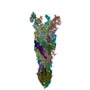
| Title | Probabilistic Structural Models of Mature P22 Bacteriophage Portal, Hub, and Tailspike proteins | ||||||||||||
 Components Components |
| ||||||||||||
 Keywords Keywords | VIRAL PROTEIN / virion / portal / tailspike / adhesin | ||||||||||||
| Function / homology |  Function and homology information Function and homology informationviral DNA genome packaging, headful / symbiont entry into host cell via disruption of host cell wall peptidoglycan / endo-1,3-alpha-L-rhamnosidase activity / symbiont entry into host cell via disruption of host cell envelope lipopolysaccharide / viral portal complex / symbiont genome ejection through host cell envelope, short tail mechanism / viral DNA genome packaging / virus tail, fiber / symbiont entry into host cell via disruption of host cell envelope / virus tail ...viral DNA genome packaging, headful / symbiont entry into host cell via disruption of host cell wall peptidoglycan / endo-1,3-alpha-L-rhamnosidase activity / symbiont entry into host cell via disruption of host cell envelope lipopolysaccharide / viral portal complex / symbiont genome ejection through host cell envelope, short tail mechanism / viral DNA genome packaging / virus tail, fiber / symbiont entry into host cell via disruption of host cell envelope / virus tail / symbiont entry into host / Hydrolases; Glycosylases; Glycosidases, i.e. enzymes that hydrolyse O- and S-glycosyl compounds / adhesion receptor-mediated virion attachment to host cell / virion assembly / hydrolase activity / virion attachment to host cell Similarity search - Function | ||||||||||||
| Biological species |  Enterobacteria phage P22 (virus) Enterobacteria phage P22 (virus) | ||||||||||||
| Method | ELECTRON MICROSCOPY / single particle reconstruction / cryo EM / Resolution: 10.5 Å | ||||||||||||
 Authors Authors | Pintilie, G. / Chen, D.H. / Haase-Pettingell, C.A. / King, J.A. / Chiu, W. | ||||||||||||
| Funding support |  United States, 3items United States, 3items
| ||||||||||||
 Citation Citation |  Journal: Biophys J / Year: 2016 Journal: Biophys J / Year: 2016Title: Resolution and Probabilistic Models of Components in CryoEM Maps of Mature P22 Bacteriophage. Authors: Grigore Pintilie / Dong-Hua Chen / Cameron A Haase-Pettingell / Jonathan A King / Wah Chiu /  Abstract: CryoEM continues to produce density maps of larger and more complex assemblies with multiple protein components of mixed symmetries. Resolution is not always uniform throughout a cryoEM map, and it ...CryoEM continues to produce density maps of larger and more complex assemblies with multiple protein components of mixed symmetries. Resolution is not always uniform throughout a cryoEM map, and it can be useful to estimate the resolution in specific molecular components of a large assembly. In this study, we present procedures to 1) estimate the resolution in subcomponents by gold-standard Fourier shell correlation (FSC); 2) validate modeling procedures, particularly at medium resolutions, which can include loop modeling and flexible fitting; and 3) build probabilistic models that combine high-accuracy priors (such as crystallographic structures) with medium-resolution cryoEM densities. As an example, we apply these methods to new cryoEM maps of the mature bacteriophage P22, reconstructed without imposing icosahedral symmetry. Resolution estimates based on gold-standard FSC show the highest resolution in the coat region (7.6 Å), whereas other components are at slightly lower resolutions: portal (9.2 Å), hub (8.5 Å), tailspike (10.9 Å), and needle (10.5 Å). These differences are indicative of inherent structural heterogeneity and/or reconstruction accuracy in different subcomponents of the map. Probabilistic models for these subcomponents provide new insights, to our knowledge, and structural information when taking into account uncertainty given the limitations of the observed density. | ||||||||||||
| History |
|
- Structure visualization
| Movie |
 Movie viewer Movie viewer |
|---|---|
| Structure viewer | Molecule:  Molmil Molmil Jmol/JSmol Jmol/JSmol |
- Downloads & links
- Download
| PDBx/mmCIF format |  5gai.cif.gz 5gai.cif.gz | 2 MB | Display |  PDBx/mmCIF format PDBx/mmCIF format |
|---|---|---|---|---|
| PDB format |  pdb5gai.ent.gz pdb5gai.ent.gz | 1.7 MB | Display |  PDB format PDB format |
| PDBx/mmJSON format |  5gai.json.gz 5gai.json.gz | Tree view |  PDBx/mmJSON format PDBx/mmJSON format | |
| Others |  Other downloads Other downloads |
-Validation report
| Arichive directory |  https://data.pdbj.org/pub/pdb/validation_reports/ga/5gai https://data.pdbj.org/pub/pdb/validation_reports/ga/5gai ftp://data.pdbj.org/pub/pdb/validation_reports/ga/5gai ftp://data.pdbj.org/pub/pdb/validation_reports/ga/5gai | HTTPS FTP |
|---|
-Related structure data
| Related structure data |  8005MC M: map data used to model this data C: citing same article ( |
|---|---|
| Similar structure data |
- Links
Links
- Assembly
| Deposited unit | 
|
|---|---|
| 1 |
|
- Components
| #1: Protein | Mass: 82397.906 Da / Num. of mol.: 12 Source method: isolated from a genetically manipulated source Source: (gene. exp.)  Enterobacteria phage P22 (virus) / Production host: Enterobacteria phage P22 (virus) / Production host:  #2: Protein | Mass: 15957.813 Da / Num. of mol.: 12 / Mutation: P150A Source method: isolated from a genetically manipulated source Source: (gene. exp.)  Enterobacteria phage P22 (virus) / Production host: Enterobacteria phage P22 (virus) / Production host:  #3: Protein | Mass: 71361.875 Da / Num. of mol.: 3 Source method: isolated from a genetically manipulated source Source: (gene. exp.)  Enterobacteria phage P22 (virus) / Production host: Enterobacteria phage P22 (virus) / Production host:  References: UniProt: P12528, Hydrolases; Glycosylases; Glycosidases, i.e. enzymes that hydrolyse O- and S-glycosyl compounds |
|---|
-Experimental details
-Experiment
| Experiment | Method: ELECTRON MICROSCOPY |
|---|---|
| EM experiment | Aggregation state: PARTICLE / 3D reconstruction method: single particle reconstruction |
- Sample preparation
| Component | Name: Enterobacteria phage P22 / Type: VIRUS / Entity ID: all / Source: NATURAL |
|---|---|
| Molecular weight | Value: 50.7 MDa / Experimental value: YES |
| Source (natural) | Organism:  Enterobacteria phage P22 (virus) / Strain: 13-am H101/C17 Enterobacteria phage P22 (virus) / Strain: 13-am H101/C17 |
| Details of virus | Empty: NO / Enveloped: NO / Isolate: STRAIN / Type: VIRION |
| Natural host | Organism: Salmonella enterica subsp. enterica serovar Typhimurium Strain: LT2 |
| Virus shell | Name: gp5 / Diameter: 710 nm / Triangulation number (T number): 7 |
| Buffer solution | pH: 7.6 |
| Buffer component | Name: 25 mM MgCl2 |
| Specimen | Embedding applied: NO / Shadowing applied: NO / Staining applied: NO / Vitrification applied: YES |
| Vitrification | Instrument: FEI VITROBOT MARK III / Cryogen name: ETHANE / Humidity: 90 % / Chamber temperature: 120 K / Details: Blot for 2 seconds before plunging. |
- Electron microscopy imaging
| Microscopy | Model: JEOL 3200FSC |
|---|---|
| Electron gun | Electron source:  FIELD EMISSION GUN / Accelerating voltage: 300 kV / Illumination mode: FLOOD BEAM FIELD EMISSION GUN / Accelerating voltage: 300 kV / Illumination mode: FLOOD BEAM |
| Electron lens | Mode: BRIGHT FIELD / Nominal magnification: 40000 X / Calibrated magnification: 70600 X / Nominal defocus max: 4000 nm / Nominal defocus min: 1000 nm / Calibrated defocus min: 1000 nm / Calibrated defocus max: 4000 nm / Cs: 4.1 mm / Alignment procedure: BASIC |
| Specimen holder | Cryogen: NITROGEN / Specimen holder model: JEOL 3200FSC CRYOHOLDER / Temperature (max): 102 K / Temperature (min): 100 K |
| Image recording | Electron dose: 25 e/Å2 / Film or detector model: GATAN ULTRASCAN 10000 (10k x 10k) / Num. of real images: 1130 Details: Every image was 2x hardware binned from Gatan 10kx10k CCD. |
| EM imaging optics | Energyfilter name: In-column Omega Filter / Energyfilter upper: 20 eV / Energyfilter lower: 0 eV |
| Image scans | Sampling size: 9 µm / Width: 5000 / Height: 5000 |
- Processing
| EM software |
| |||||||||||||||||||||||||||||||||||
|---|---|---|---|---|---|---|---|---|---|---|---|---|---|---|---|---|---|---|---|---|---|---|---|---|---|---|---|---|---|---|---|---|---|---|---|---|
| CTF correction | Type: PHASE FLIPPING AND AMPLITUDE CORRECTION | |||||||||||||||||||||||||||||||||||
| Particle selection | Num. of particles selected: 79731 | |||||||||||||||||||||||||||||||||||
| Symmetry | Point symmetry: C1 (asymmetric) | |||||||||||||||||||||||||||||||||||
| 3D reconstruction | Resolution: 10.5 Å / Resolution method: FSC 0.143 CUT-OFF / Num. of particles: 79731 / Algorithm: FOURIER SPACE / Symmetry type: POINT |
 Movie
Movie Controller


 PDBj
PDBj