 Movie
Movie Controller

[English] 日本語
 Yorodumi
- EMDB-12985: Low resolution reconstruction of the dATP-inhibited complex of th... -
+ Open data

- Basic information
| Entry | Database: EMDB / ID: EMD-12985 | |||||||||||||||
|---|---|---|---|---|---|---|---|---|---|---|---|---|---|---|---|---|
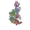
| Title | Low resolution reconstruction of the dATP-inhibited complex of the ribonucleotide reductase NrdA and NrdB proteins from Leeuwenhoekiella blandensis | |||||||||||||||
 Map data Map data | ||||||||||||||||
 Sample Sample |
| |||||||||||||||
| Biological species |  Leeuwenhoekiella blandensis (bacteria) Leeuwenhoekiella blandensis (bacteria) | |||||||||||||||
| Method | single particle reconstruction / cryo EM / Resolution: 8.2 Å | |||||||||||||||
 Authors Authors | Banerjee I / Rozman Grinberg I / Sjoberg BM / Logan DT | |||||||||||||||
| Funding support |  Sweden, 4 items Sweden, 4 items
| |||||||||||||||
 Citation Citation | Journal: Front Mol Biosci / Year: 2021 Title: Solution Structure of the dATP-Inactivated Class I Ribonucleotide Reductase From by SAXS and Cryo-Electron Microscopy. Authors: Mahmudul Hasan / Ipsita Banerjee / Inna Rozman Grinberg / Britt-Marie Sjöberg / Derek T Logan / Abstract: The essential enzyme ribonucleotide reductase (RNR) is highly regulated both at the level of overall activity and substrate specificity. Studies of class I, aerobic RNRs have shown that overall ...The essential enzyme ribonucleotide reductase (RNR) is highly regulated both at the level of overall activity and substrate specificity. Studies of class I, aerobic RNRs have shown that overall activity is downregulated by the binding of dATP to a small domain known as the ATP-cone often found at the N-terminus of RNR subunits, causing oligomerization that prevents formation of a necessary αβ complex between the catalytic (α) and radical generating (β) subunits. In some relatively rare organisms with RNRs of the subclass NrdAi, the ATP-cone is found at the N-terminus of the β subunit rather than more commonly the α subunit. Binding of dATP to the ATP-cone in β results in formation of an unusual β tetramer. However, the structural basis for how the formation of the active complex is hindered by such oligomerization has not been studied. Here we analyse the low-resolution three-dimensional structures of the separate subunits of an RNR from subclass NrdAi, as well as the αβ octamer that forms in the presence of dATP. The results reveal a type of oligomer not previously seen for any class of RNR and suggest a mechanism for how binding of dATP to the ATP-cone switches off catalysis by sterically preventing formation of the asymmetrical αβ complex. | |||||||||||||||
| History |
|
- Structure visualization
| Movie |
 Movie viewer Movie viewer |
|---|---|
| Structure viewer | EM map: SurfViewMolmilJmol/JSmol |
| Supplemental images |

- Downloads & links
-EMDB archive
| Map data | emd_12985.map.gz | 322.8 MB | EMDB map data format | |
|---|---|---|---|---|
| Header (meta data) | emd-12985-v30.xmlemd-12985.xml | 18 KB 18 KB | Display Display | EMDB header |
| FSC (resolution estimation) | emd_12985_fsc.xml | 15.7 KB | Display | FSC data file |
| Images |  emd_12985.png emd_12985.png | 119.4 KB | ||
| Masks | emd_12985_msk_1.map | 343 MB | Mask map | |
| Archive directory |  http://ftp.pdbj.org/pub/emdb/structures/EMD-12985ftp://ftp.pdbj.org/pub/emdb/structures/EMD-12985 http://ftp.pdbj.org/pub/emdb/structures/EMD-12985ftp://ftp.pdbj.org/pub/emdb/structures/EMD-12985 | HTTPS FTP |
-Related structure data
| Similar structure data |
|---|


-Links
| EMDB pages | EMDB (EBI/PDBe) / EMDataResource |
|---|
-Map
| File | Download / File: emd_12985.map.gz / Format: CCP4 / Size: 343 MB / Type: IMAGE STORED AS FLOATING POINT NUMBER (4 BYTES) | ||||||||||||||||||||||||||||||||||||||||||||||||||||||||||||||||||||
|---|---|---|---|---|---|---|---|---|---|---|---|---|---|---|---|---|---|---|---|---|---|---|---|---|---|---|---|---|---|---|---|---|---|---|---|---|---|---|---|---|---|---|---|---|---|---|---|---|---|---|---|---|---|---|---|---|---|---|---|---|---|---|---|---|---|---|---|---|---|
| Projections & slices | Image control
Images are generated by Spider. | ||||||||||||||||||||||||||||||||||||||||||||||||||||||||||||||||||||
| Voxel size | X=Y=Z: 0.82 Å | ||||||||||||||||||||||||||||||||||||||||||||||||||||||||||||||||||||
| Density |
| ||||||||||||||||||||||||||||||||||||||||||||||||||||||||||||||||||||
| Symmetry | Space group: 1 | ||||||||||||||||||||||||||||||||||||||||||||||||||||||||||||||||||||
| Details | EMDB XML:
CCP4 map header:
| ||||||||||||||||||||||||||||||||||||||||||||||||||||||||||||||||||||
 Z (Sec.)
 Y (Row.)
 X (Col.)


-Supplemental data
-Mask #1
| File | emd_12985_msk_1.map | ||||||||||||
|---|---|---|---|---|---|---|---|---|---|---|---|---|---|
| Projections & Slices |
| ||||||||||||
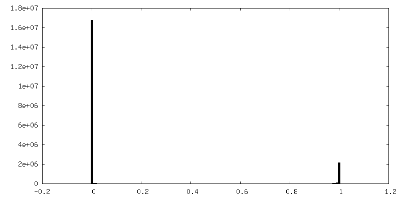
| Density Histograms |


- Sample components
-Entire : dATP-inhibited complex of NrdA and NrdB subunits from Leeuwenhoek...
| Entire | Name: dATP-inhibited complex of NrdA and NrdB subunits from Leeuwenhoekiella blandensis |
|---|---|
| Components |
|
-Supramolecule #1: dATP-inhibited complex of NrdA and NrdB subunits from Leeuwenhoek...
| Supramolecule | Name: dATP-inhibited complex of NrdA and NrdB subunits from Leeuwenhoekiella blandensis type: complex / ID: 1 / Parent: 0 / Macromolecule list: all Details: The complex was formed by incubating the NrdA and NrdB components in the presence of 2mM dATP followed by glutaraldehyde crosslinking on a gel filtration column. |
|---|---|
| Source (natural) | Organism: Leeuwenhoekiella blandensis (bacteria) |
| Molecular weight | Theoretical: 490 KDa |
-Macromolecule #1: Ribonucleotide reductase catalytic subunit from Leeuwenhoekiella ...
| Macromolecule | Name: Ribonucleotide reductase catalytic subunit from Leeuwenhoekiella blandensis type: protein_or_peptide / ID: 1 / Enantiomer: LEVO / EC number: ribonucleoside-diphosphate reductase |
|---|---|
| Source (natural) | Organism: Leeuwenhoekiella blandensis (bacteria) |
| Recombinant expression | Organism: |
| Sequence | String: MGSSHHHHHH SSGLVPRGSH MRENTTKLEN STETNVQHTK DNDLLKARRD ALQDATEKNT DEPSFDWLTE HSRSFLAAGY LSEGVSAEER IREIADRAE EILRMPGFSD KFYKYMGEGY FSLASPVWSN FGKKRGLPIS CFGSHIDDDM GNILYTQSEV G MMSKLGGG ...String: MGSSHHHHHH SSGLVPRGSH MRENTTKLEN STETNVQHTK DNDLLKARRD ALQDATEKNT DEPSFDWLTE HSRSFLAAGY LSEGVSAEER IREIADRAE EILRMPGFSD KFYKYMGEGY FSLASPVWSN FGKKRGLPIS CFGSHIDDDM GNILYTQSEV G MMSKLGGG TSGYFGKIRH RGAAVKNNGY ASGAVHIMQL FDKMVDVVSQ GSVRRGRFSP YLPISHPDIK EF LEIGTEG NSIQQLTHGV TVDSTWMQEM IDGDTDKREV WAKVLQRRGE MGYPYIFYTD NANNGKPDVY KDK GHDIYA SNLCTEIMLP SSDEWSFVCV LSSINVLHYD KWKNTDAVET MVCFLDAVLT EFIDKLEEYR DSDN RDHRQ TFMFMERAYN FAKSNRALGL GVLGWHSLLQ SKRHAFDSQE AYDLNSEIFR EIKQRSYKAS EELAE KFGE PETLKGYGRR NATLNAIAPT TSSAFILGQV SQGIEPIWSN VYVKDIAKIK TTIKNPFLEE LFEEKG MNT PEVWRSVRDN DGSVQHLEFL TEQEKDVFKT YAEIDQMAII YQAANRQNHI DQGQSINLLV HPDMPIK EI NKIHITAWKL GLKSLYYQHS MNAAQKFKQK KECVSCEA |
-Macromolecule #2: Ribonucleotide reductase radical generating subunit from Leeuwenh...
| Macromolecule | Name: Ribonucleotide reductase radical generating subunit from Leeuwenhoekiella blandensis type: protein_or_peptide / ID: 2 / Enantiomer: LEVO |
|---|---|
| Source (natural) | Organism: Leeuwenhoekiella blandensis (bacteria) |
| Recombinant expression | Organism: |
| Sequence | String: MSSQEIKKII KRDYSTAPFV LEKITNAIAN AMAALGHGSE QDAKLISMQV YESLLNNKEQ ESEYIPTVE QVQDMVEDKL MSSEFHDVAK AYIIYRNKRA LERKTNIFEK RINLKPYEYP E LNEYVAAI RHSYWIHTEF NFTSDIQDFK TGLSEVERSA IKNTMLAISQ ...String: MSSQEIKKII KRDYSTAPFV LEKITNAIAN AMAALGHGSE QDAKLISMQV YESLLNNKEQ ESEYIPTVE QVQDMVEDKL MSSEFHDVAK AYIIYRNKRA LERKTNIFEK RINLKPYEYP E LNEYVAAI RHSYWIHTEF NFTSDIQDFK TGLSEVERSA IKNTMLAISQ IEVAVKTFWG DV HHRLPKP EIAAVGATFA ESEVRHHDAY SHLLEILGLN EEFKELKKKP VIMKRVHYLE TSL KHAKSD DDREYTESIL LFALFIEHVS LFSQFLIIMA FNKHKNMLKG ISNAVEATSK EEQI HGDFG VDIINIIKKE NPEWFDEEHN NLIKEMCLNS FEAESKVVDW IFEKGELDFL PKAVI NEFL KNRFNKSLEA IGLEKLFDID EALLQETEWF DDEIIGTKHG DFFVKRSINY SKRTQS ITS DDLF |
-Experimental details
-Structure determination
| Method | cryo EM |
|---|---|
 Processing Processing | single particle reconstruction |
| Aggregation state | particle |
-Sample preparation
| Buffer | pH: 7.6 Component:
| |||||||||||||||||||||
|---|---|---|---|---|---|---|---|---|---|---|---|---|---|---|---|---|---|---|---|---|---|---|
| Grid | Model: Quantifoil R1.2/1.3 / Material: COPPER / Pretreatment - Type: GLOW DISCHARGE | |||||||||||||||||||||
| Vitrification | Cryogen name: ETHANE / Chamber humidity: 100 % / Chamber temperature: 277 K / Instrument: FEI VITROBOT MARK IV Details: Blot time 5 s, blot force -5, wait time 1 s, drain time 0 s. | |||||||||||||||||||||
| Details | The specimen was taken directly from a fraction of a size exclusion chromatography run done in the presence of glutaraldehyde. |
- Electron microscopy
| Microscope | FEI TITAN KRIOS |
|---|---|
| Image recording | Film or detector model: GATAN K2 QUANTUM (4k x 4k) / Detector mode: COUNTING / Digitization - Dimensions - Width: 4000 pixel / Digitization - Dimensions - Height: 4000 pixel / Digitization - Frames/image: 1-40 / Number grids imaged: 1 / Number real images: 493 / Average exposure time: 4.0 sec. / Average electron dose: 47.1 e/Å2 Details: Data recorded at the SciLifeLab facility in Umea, Sweden |
| Electron beam | Acceleration voltage: 300 kV / Electron source:  FIELD EMISSION GUN FIELD EMISSION GUN |
| Electron optics | C2 aperture diameter: 70.0 µm / Illumination mode: SPOT SCAN / Imaging mode: BRIGHT FIELD / Cs: 2.7 mm / Nominal defocus max: -0.003 µm / Nominal defocus min: -0.0015 µm / Nominal magnification: 165000 |
| Sample stage | Specimen holder model: FEI TITAN KRIOS AUTOGRID HOLDER / Cooling holder cryogen: NITROGEN |
| Experimental equipment |  Model: Titan Krios / Image courtesy: FEI Company |


