DnaA-Dps complex / Oxidoreductases; Oxidizing metal ions / oxidoreductase activity, acting on metal ions / nucleoid / chromosome condensation / response to starvation / response to stress / ferric iron binding / negative regulation of DNA-templated DNA replication initiation / intracellular iron ion homeostasis ...DnaA-Dps complex / Oxidoreductases; Oxidizing metal ions / oxidoreductase activity, acting on metal ions / nucleoid / chromosome condensation / response to starvation / response to stress / ferric iron binding / negative regulation of DNA-templated DNA replication initiation / intracellular iron ion homeostasis / DNA binding / membrane / identical protein binding / cytoplasm Similarity search - Function
DNA protection during starvation protein, gammaproteobacteria / Dps protein family signature 2. / Dps protein family signature 1. / DNA-binding protein Dps, conserved site / DNA-binding protein Dps / Ferritin/DPS protein domain / Ferritin-like domain / Ferritin-like / Ferritin-like superfamily Similarity search - Domain/homology
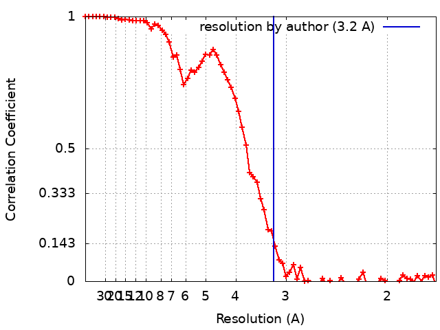
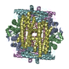
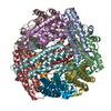
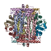
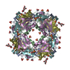
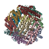
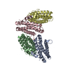
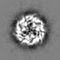
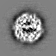
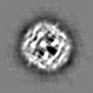
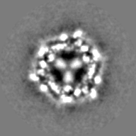
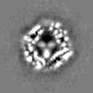
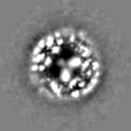
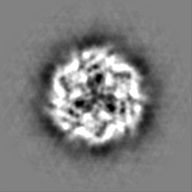
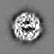
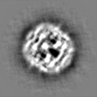
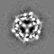
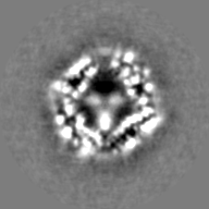
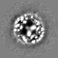
Journal: Int J Mol Sci / Year: 2021 Title: Structural Rearrangement of Dps-DNA Complex Caused by Divalent Mg and Fe Cations. Authors: Liubov Dadinova / Roman Kamyshinsky / Yury Chesnokov / Andrey Mozhaev / Vladimir Matveev / Andrey Gruzinov / Alexander Vasiliev / Eleonora Shtykova / Abstract: Two independent, complementary methods of structural analysis were used to elucidate the effect of divalent magnesium and iron cations on the structure of the protective Dps-DNA complex. Small-angle ...Two independent, complementary methods of structural analysis were used to elucidate the effect of divalent magnesium and iron cations on the structure of the protective Dps-DNA complex. Small-angle X-ray scattering (SAXS) and cryo-electron microscopy (cryo-EM) demonstrate that Mg ions block the N-terminals of the Dps protein preventing its interaction with DNA. Non-interacting macromolecules of Dps and DNA remain in the solution in this case. The subsequent addition of the chelating agent (EDTA) leads to a complete restoration of the structure of the complex. Different effect was observed when Fe cations were added to the Dps-DNA complex; the presence of Fe in solution leads to the total complex destruction and aggregation without possibility of the complex restoration with the chelating agent. Here, we discuss these different responses of the Dps-DNA complex on the presence of additional free metal cations, investigating the structure of the Dps protein with and without cations using SAXS and cryo-EM. Additionally, the single particle analysis of Dps with accumulated iron performed by cryo-EM shows localization of iron nanoparticles inside the Dps cavity next to the acidic (hydrophobic) pore, near three glutamate residues.
History
Deposition
May 17, 2021
-
Header (metadata) release
Jun 16, 2021
-
Map release
Jun 16, 2021
-
Update
Jul 14, 2021
-
Current status
Jul 14, 2021
Processing site: PDBe / Status: Released
-
Structure visualization
Movie
Surface view with section colored by density value
In the structure databanks used in Yorodumi, some data are registered as the other names, "COVID-19 virus" and "2019-nCoV". Here are the details of the virus and the list of structure data.
Jan 31, 2019. EMDB accession codes are about to change! (news from PDBe EMDB page)
EMDB accession codes are about to change! (news from PDBe EMDB page)
The allocation of 4 digits for EMDB accession codes will soon come to an end. Whilst these codes will remain in use, new EMDB accession codes will include an additional digit and will expand incrementally as the available range of codes is exhausted. The current 4-digit format prefixed with “EMD-” (i.e. EMD-XXXX) will advance to a 5-digit format (i.e. EMD-XXXXX), and so on. It is currently estimated that the 4-digit codes will be depleted around Spring 2019, at which point the 5-digit format will come into force.
The EM Navigator/Yorodumi systems omit the EMD- prefix.
Related info.:Q: What is EMD? / ID/Accession-code notation in Yorodumi/EM Navigator
Yorodumi is a browser for structure data from EMDB, PDB, SASBDB, etc.
This page is also the successor to EM Navigator detail page, and also detail information page/front-end page for Omokage search.
The word "yorodu" (or yorozu) is an old Japanese word meaning "ten thousand". "mi" (miru) is to see.
Related info.:EMDB / PDB / SASBDB / Comparison of 3 databanks / Yorodumi Search / Aug 31, 2016. New EM Navigator & Yorodumi / Yorodumi Papers / Jmol/JSmol / Function and homology information / Changes in new EM Navigator and Yorodumi
 Movie
Movie Controller


 Open data
 Basic information
 Map data
 Sample
 Function and homology information
 Authors
 Russian Federation, 1 items
  Citation
 Structure visualization
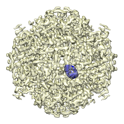
 Downloads & links
 emd_12961.png
 http://ftp.pdbj.org/pub/emdb/structures/EMD-12961


 Z (Sec.)
 Y (Row.)
 X (Col.)


 Sample components
 Processing
 Electron microscopy
 FIELD EMISSION GUN