 Movie
Movie Controller


+ Open data

- Basic information
| Entry | Database: EMDB / ID: EMD-10161 | |||||||||
|---|---|---|---|---|---|---|---|---|---|---|
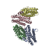
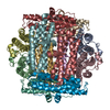
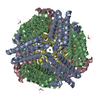
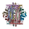
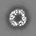
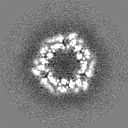
| Title | Structure of DPS determined at 100 keV | |||||||||
 Map data Map data | Masked and sharpened (B = -132 A^2) DPS map | |||||||||
 Sample Sample |
| |||||||||
| Biological species |  | |||||||||
| Method | single particle reconstruction / cryo EM / Resolution: 3.4 Å | |||||||||
 Authors Authors | Naydenova K / McMullan G / Peet MJ / Lee Y / Edwards PC / Chen S / Leahy E / Henderson R / Russo CJ | |||||||||
| Funding support |  United Kingdom, 2 items United Kingdom, 2 items
| |||||||||
 Citation Citation | Journal: IUCrJ / Year: 2019 Title: CryoEM at 100 keV: a demonstration and prospects. Authors: K Naydenova / G McMullan / M J Peet / Y Lee / P C Edwards / S Chen / E Leahy / S Scotcher / R Henderson / C J Russo / Abstract: 100 kV is investigated as the operating voltage for single-particle electron cryomicroscopy (cryoEM). Reducing the electron energy from the current standard of 300 or 200 keV offers both cost ...100 kV is investigated as the operating voltage for single-particle electron cryomicroscopy (cryoEM). Reducing the electron energy from the current standard of 300 or 200 keV offers both cost savings and potentially improved imaging. The latter follows from recent measurements of radiation damage to biological specimens by high-energy electrons, which show that at lower energies there is an increased amount of information available per unit damage. For frozen hydrated specimens around 300 Å in thickness, the predicted optimal electron energy for imaging is 100 keV. Currently available electron cryomicroscopes in the 100-120 keV range are not optimized for cryoEM as they lack both the spatially coherent illumination needed for the high defocus used in cryoEM and imaging detectors optimized for 100 keV electrons. To demonstrate the potential of imaging at 100 kV, the voltage of a standard, commercial 200 kV field-emission gun (FEG) microscope was reduced to 100 kV and a side-entry cryoholder was used. As high-efficiency, large-area cameras are not currently available for 100 keV electrons, a commercial hybrid pixel camera designed for X-ray detection was attached to the camera chamber and was used for low-dose data collection. Using this configuration, five single-particle specimens were imaged: hepatitis B virus capsid, bacterial 70S ribosome, catalase, DNA protection during starvation protein and haemoglobin, ranging in size from 4.5 MDa to 64 kDa with corresponding diameters from 320 to 72 Å. These five data sets were used to reconstruct 3D structures with resolutions between 8.4 and 3.4 Å. Based on this work, the practical advantages and current technological limitations to single-particle cryoEM at 100 keV are considered. These results are also discussed in the context of future microscope development towards the goal of rapid, simple and widely available structure determination of any purified biological specimen. | |||||||||
| History |
|
- Structure visualization
| Movie |
 Movie viewer Movie viewer |
|---|---|
| Structure viewer | EM map: SurfViewMolmilJmol/JSmol |
| Supplemental images |

- Downloads & links
-EMDB archive
| Map data | emd_10161.map.gz | 1 MB | EMDB map data format | |
|---|---|---|---|---|
| Header (meta data) | emd-10161-v30.xmlemd-10161.xml | 14.8 KB 14.8 KB | Display Display | EMDB header |
| FSC (resolution estimation) | emd_10161_fsc.xml | 4.6 KB | Display | FSC data file |
| Images | 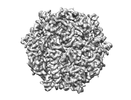 emd_10161.png emd_10161.png | 161.4 KB | ||
| Masks | emd_10161_msk_1.map | 8 MB | Mask map | |
| Others | emd_10161_additional.map.gzemd_10161_additional_1.map.gzemd_10161_half_map_1.map.gzemd_10161_half_map_2.map.gz | 6 MB 6 MB 6 MB 6 MB | ||
| Archive directory |  http://ftp.pdbj.org/pub/emdb/structures/EMD-10161ftp://ftp.pdbj.org/pub/emdb/structures/EMD-10161 http://ftp.pdbj.org/pub/emdb/structures/EMD-10161ftp://ftp.pdbj.org/pub/emdb/structures/EMD-10161 | HTTPS FTP |
-Related structure data
| Related structure data | C: citing same article ( |
|---|---|
| Similar structure data | |
| EM raw data | EMPIAR-10297 (Title: Micrographs of DPS collected at 100 keV using a hybrid pixel direct electron detector Data size: 23.3 Data #1: Raw multi-frame micrographs of DPS recorded at 100 keV [micrographs - multiframe]) |


-Links
| EMDB pages | EMDB (EBI/PDBe) / EMDataResource |
|---|
-Map
| File | Download / File: emd_10161.map.gz / Format: CCP4 / Size: 8 MB / Type: IMAGE STORED AS FLOATING POINT NUMBER (4 BYTES) | ||||||||||||||||||||||||||||||||||||||||||||||||||||||||||||
|---|---|---|---|---|---|---|---|---|---|---|---|---|---|---|---|---|---|---|---|---|---|---|---|---|---|---|---|---|---|---|---|---|---|---|---|---|---|---|---|---|---|---|---|---|---|---|---|---|---|---|---|---|---|---|---|---|---|---|---|---|---|
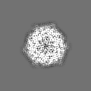
| Annotation | Masked and sharpened (B = -132 A^2) DPS map | ||||||||||||||||||||||||||||||||||||||||||||||||||||||||||||
| Projections & slices | Image control
Images are generated by Spider. | ||||||||||||||||||||||||||||||||||||||||||||||||||||||||||||
| Voxel size | X=Y=Z: 1.47 Å | ||||||||||||||||||||||||||||||||||||||||||||||||||||||||||||
| Density |
| ||||||||||||||||||||||||||||||||||||||||||||||||||||||||||||
| Symmetry | Space group: 1 | ||||||||||||||||||||||||||||||||||||||||||||||||||||||||||||
| Details | EMDB XML:
CCP4 map header:
| ||||||||||||||||||||||||||||||||||||||||||||||||||||||||||||
 Z (Sec.)
 Y (Row.)
 X (Col.)


-Supplemental data
-Mask #1
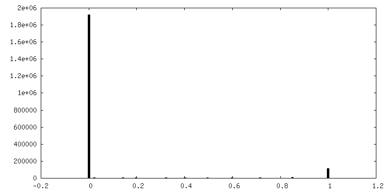
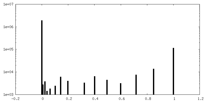
| File | emd_10161_msk_1.map | ||||||||||||
|---|---|---|---|---|---|---|---|---|---|---|---|---|---|
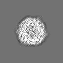
| Projections & Slices |
| ||||||||||||
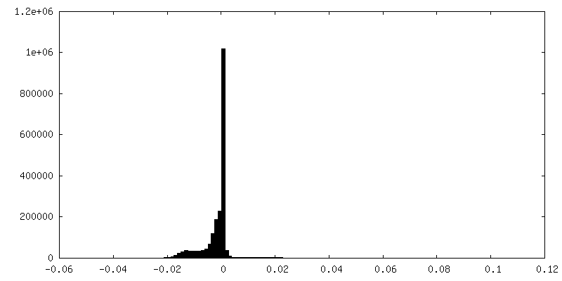
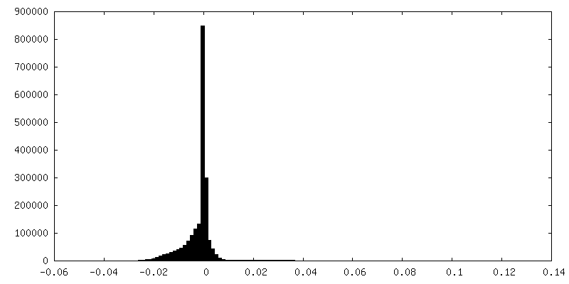
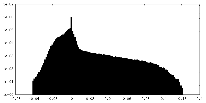
| Density Histograms |


-Additional map: Unsharpened DPS map
| File | emd_10161_additional.map | ||||||||||||
|---|---|---|---|---|---|---|---|---|---|---|---|---|---|
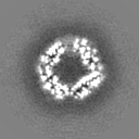
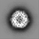
| Annotation | Unsharpened DPS map | ||||||||||||
| Projections & Slices |
| ||||||||||||
| Density Histograms |


-Additional map: Unsharpened DPS map
| File | emd_10161_additional_1.map | ||||||||||||
|---|---|---|---|---|---|---|---|---|---|---|---|---|---|
| Annotation | Unsharpened DPS map | ||||||||||||
| Projections & Slices |
| ||||||||||||
| Density Histograms |


-Half map: DPS half map 1
| File | emd_10161_half_map_1.map | ||||||||||||
|---|---|---|---|---|---|---|---|---|---|---|---|---|---|
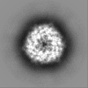
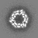
| Annotation | DPS half map 1 | ||||||||||||
| Projections & Slices |
| ||||||||||||
| Density Histograms |


-Half map: DPS half map 2
| File | emd_10161_half_map_2.map | ||||||||||||
|---|---|---|---|---|---|---|---|---|---|---|---|---|---|
| Annotation | DPS half map 2 | ||||||||||||
| Projections & Slices |
| ||||||||||||
| Density Histograms |


- Sample components
-Entire : DPS from E. coli
| Entire | Name: DPS from E. coli |
|---|---|
| Components |
|
-Supramolecule #1: DPS from E. coli
| Supramolecule | Name: DPS from E. coli / type: complex / ID: 1 / Parent: 0 |
|---|---|
| Source (natural) | Organism: |
| Recombinant expression | Organism: |
-Experimental details
-Structure determination
| Method | cryo EM |
|---|---|
 Processing Processing | single particle reconstruction |
| Aggregation state | particle |
-Sample preparation
| Buffer | pH: 7.7 |
|---|---|
| Grid | Model: UltrAuFoil / Material: GOLD / Mesh: 300 |
| Vitrification | Cryogen name: ETHANE |
- Electron microscopy
| Microscope | FEI TECNAI F20 |
|---|---|
| Image recording | Film or detector model: OTHER / Average electron dose: 24.64 e/Å2 |
| Electron beam | Acceleration voltage: 100 kV / Electron source:  FIELD EMISSION GUN FIELD EMISSION GUN |
| Electron optics | Illumination mode: FLOOD BEAM / Imaging mode: BRIGHT FIELD |
| Experimental equipment |  Model: Tecnai F20 / Image courtesy: FEI Company |
-Image processing
| Final reconstruction | Applied symmetry - Point group: T (tetrahedral) / Resolution.type: BY AUTHOR / Resolution: 3.4 Å / Resolution method: FSC 0.143 CUT-OFF / Number images used: 16507 |
|---|---|
| Initial angle assignment | Type: MAXIMUM LIKELIHOOD |
| Final angle assignment | Type: MAXIMUM LIKELIHOOD |
| FSC plot (resolution estimation) |  |
