- EMDB-12757: Rabbit 80S ribosome stalled close to the mutated SARS-CoV-2 slipp... -
+
Open data
ID or keywords:
Loading...
-
Basic information
Entry
Database: EMDB / ID: EMD-12757
Title
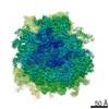
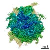
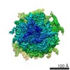
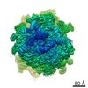
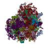
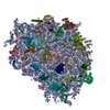
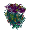
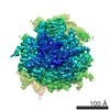
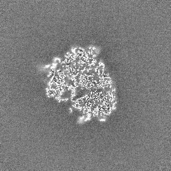
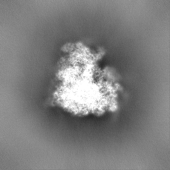
Rabbit 80S ribosome stalled close to the mutated SARS-CoV-2 slippery site by a pseudoknot (classified for pseudoknot)
Map data
Sample
Complex: Rabbit 80S ribosome stalled close to the mutated SARS-CoV-2 slippery site by a pseudoknot
Complex: Rabbit 80S ribosome stalled
RNA: x 4 types
Protein or peptide: x 79 types
Complex: SARS-CoV-2 slippery site by a pseudoknot
RNA: x 1 types
Complex: Rabbit 80S ribosome stalled
RNA: x 2 types
Complex: Replicase polyprotein 1ab
Ligand: x 6 types
Keywords
Frameshift / virus / pseudoknot / RIBOSOME
Function / homology
Function and homology information
Major pathway of rRNA processing in the nucleolus and cytosol / GTP hydrolysis and joining of the 60S ribosomal subunit / L13a-mediated translational silencing of Ceruloplasmin expression / SRP-dependent cotranslational protein targeting to membrane / Formation of a pool of free 40S subunits / Nonsense Mediated Decay (NMD) independent of the Exon Junction Complex (EJC) / Nonsense Mediated Decay (NMD) enhanced by the Exon Junction Complex (EJC) / laminin receptor activity / 90S preribosome / ubiquitin ligase inhibitor activity ...Major pathway of rRNA processing in the nucleolus and cytosol / GTP hydrolysis and joining of the 60S ribosomal subunit / L13a-mediated translational silencing of Ceruloplasmin expression / SRP-dependent cotranslational protein targeting to membrane / Formation of a pool of free 40S subunits / Nonsense Mediated Decay (NMD) independent of the Exon Junction Complex (EJC) / Nonsense Mediated Decay (NMD) enhanced by the Exon Junction Complex (EJC) / laminin receptor activity / 90S preribosome / ubiquitin ligase inhibitor activity / positive regulation of signal transduction by p53 class mediator / phagocytic cup / protein-RNA complex assembly / translation regulator activity / rough endoplasmic reticulum / ribosomal small subunit export from nucleus / laminin binding / gastrulation / MDM2/MDM4 family protein binding / class I DNA-(apurinic or apyrimidinic site) endonuclease activity / cytosolic ribosome / DNA-(apurinic or apyrimidinic site) lyase / ribosomal large subunit biogenesis / maturation of LSU-rRNA from tricistronic rRNA transcript (SSU-rRNA, 5.8S rRNA, LSU-rRNA) / maturation of SSU-rRNA from tricistronic rRNA transcript (SSU-rRNA, 5.8S rRNA, LSU-rRNA) / positive regulation of apoptotic signaling pathway / maturation of SSU-rRNA / small-subunit processome / spindle / cytoplasmic ribonucleoprotein granule / rRNA processing / rhythmic process / regulation of translation / positive regulation of canonical Wnt signaling pathway / antimicrobial humoral immune response mediated by antimicrobial peptide / large ribosomal subunit / ribosomal small subunit assembly / virus receptor activity / protein guanylyltransferase activity / RNA endonuclease activity producing 3'-phosphomonoesters, hydrolytic mechanism / ribosome binding / 5'-3' RNA helicase activity / ribosomal small subunit biogenesis / Lyases; Phosphorus-oxygen lyases / 5S rRNA binding / ribosomal large subunit assembly / small ribosomal subunit / Assembly of the SARS-CoV-2 Replication-Transcription Complex (RTC) / symbiont-mediated suppression of host cytoplasmic pattern recognition receptor signaling pathway via inhibition of TBK1 activity / small ribosomal subunit rRNA binding / Maturation of replicase proteins / TRAF3-dependent IRF activation pathway / ISG15-specific peptidase activity / Transcription of SARS-CoV-2 sgRNAs / cytosolic small ribosomal subunit / large ribosomal subunit rRNA binding / snRNP Assembly / Translation of Replicase and Assembly of the Replication Transcription Complex / Replication of the SARS-CoV-2 genome / Hydrolases; Acting on ester bonds; Exoribonucleases producing 5'-phosphomonoesters / double membrane vesicle viral factory outer membrane / SARS coronavirus main proteinase / host cell endoplasmic reticulum-Golgi intermediate compartment / host cell endosome / 3'-5'-RNA exonuclease activity / symbiont-mediated degradation of host mRNA / 5'-3' DNA helicase activity / mRNA guanylyltransferase / symbiont-mediated suppression of host toll-like receptor signaling pathway / killing of cells of another organism / symbiont-mediated suppression of host ISG15-protein conjugation / G-quadruplex RNA binding / mRNA guanylyltransferase activity / symbiont-mediated suppression of host cytoplasmic pattern recognition receptor signaling pathway via inhibition of IRF3 activity / cytosolic large ribosomal subunit / omega peptidase activity / mRNA (guanine-N7)-methyltransferase / methyltransferase cap1 / defense response to Gram-negative bacterium / DNA helicase / symbiont-mediated suppression of host NF-kappaB cascade / SARS-CoV-2 modulates host translation machinery / symbiont-mediated perturbation of host ubiquitin-like protein modification / host cell Golgi apparatus / perikaryon / methyltransferase cap1 activity / cell differentiation / cytoplasmic translation / mRNA 5'-cap (guanine-N7-)-methyltransferase activity / cysteine-type deubiquitinase activity / ubiquitinyl hydrolase 1 / tRNA binding / Hydrolases; Acting on peptide bonds (peptidases); Cysteine endopeptidases / lyase activity / single-stranded RNA binding / mitochondrial inner membrane / postsynaptic density / rRNA binding / viral protein processing / host cell perinuclear region of cytoplasm Similarity search - Function
60s Acidic ribosomal protein / 60S acidic ribosomal protein P0 / : / 40S ribosomal protein SA / Ribosomal protein L6, N-terminal / Ribosomal protein L6, N-terminal domain / 40S ribosomal protein SA, C-terminal domain / 40S ribosomal protein SA C-terminus / Ubiquitin-like protein FUBI / 50S ribosomal protein L10, insertion domain superfamily ...60s Acidic ribosomal protein / 60S acidic ribosomal protein P0 / : / 40S ribosomal protein SA / Ribosomal protein L6, N-terminal / Ribosomal protein L6, N-terminal domain / 40S ribosomal protein SA, C-terminal domain / 40S ribosomal protein SA C-terminus / Ubiquitin-like protein FUBI / 50S ribosomal protein L10, insertion domain superfamily / 60S ribosomal protein L10P, insertion domain / Insertion domain in 60S ribosomal protein L10P / Ribosomal protein L30e / Ribosomal L15/L27a, N-terminal / : / Ribosomal protein L23 / Ribosomal protein L1, conserved site / Ribosomal protein L1 signature. / Ribosomal protein L1 / metallochaperone-like domain / TRASH domain / Ribosomal protein L1, 3-layer alpha/beta-sandwich / Ribosomal protein L41 / Ribosomal protein L41 / : / Ribosomal protein S12e signature. / Ribosomal protein S12e / Ribosomal protein L29e / Ribosomal L29e protein family / Ribosomal protein L1-like / Ribosomal protein L1/ribosomal biogenesis protein / Ribosomal protein L1p/L10e family / Ribosomal protein L27e, conserved site / Ribosomal protein L27e signature. / Small (40S) ribosomal subunit Asc1/RACK1 / Ribosomal protein L38e / Ribosomal protein L38e superfamily / Ribosomal L38e protein family / Ribosomal protein S2, eukaryotic / Ribosomal protein L19, eukaryotic / : / Ribosomal protein L10e, conserved site / Ribosomal protein L6e signature. / Ribosomal protein L10e signature. / Ribosomal protein L19/L19e conserved site / Ribosomal protein L19e signature. / Ribosomal protein L44e signature. / 40S Ribosomal protein S10 / Ribosomal protein L10e / Ribosomal protein L24e, conserved site / Ribosomal protein L24e signature. / S27a-like superfamily / Ribosomal protein L34e, conserved site / Ribosomal protein L34e signature. / Plectin/S10, N-terminal / Plectin/S10 domain / Ribosomal protein L5 eukaryotic, C-terminal / Ribosomal L18 C-terminal region / Ribosomal protein L23/L25, N-terminal / Ribosomal protein L23, N-terminal domain / : / Ribosomal protein L36e signature. / Ribosomal protein S30 / Ribosomal protein S30 / Ribosomal protein L30e signature 1. / Ribosomal protein L44e / Ribosomal protein S10, eukaryotic/archaeal / Ribosomal protein L44 / Ribosomal protein S25 / Ribosomal protein S8e subdomain, eukaryotes / S25 ribosomal protein / : / Ribosomal protein L35Ae, conserved site / Ribosomal protein L35Ae signature. / Eukaryotic Ribosomal Protein L27, KOW domain / Ribosomal protein S7e signature. / Ribosomal protein L27e / Ribosomal protein L27e superfamily / Ribosomal L27e protein family / : / Ribosomal protein S17e, conserved site / Ribosomal Protein L6, KOW domain / Ribosomal protein S17e signature. / Ribosomal protein S27a / Ribosomal protein S2, eukaryotic/archaeal / Ribosomal protein L39e, conserved site / Ribosomal protein S27a / Ribosomal protein L39e signature. / Ribosomal protein S27a / 60S ribosomal protein L35 / Ribosomal protein L30e signature 2. / Ribosomal protein L30e, conserved site / : / Ribosomal protein S3Ae, conserved site / Ribosomal protein S3Ae signature. / Ribosomal protein L34Ae / Ribosomal protein L34e / 60S ribosomal protein L19 / Ribosomal protein L6e / 40S ribosomal protein S29/30S ribosomal protein S14 type Z Similarity search - Domain/homology
Small ribosomal subunit protein eS32 / Large ribosomal subunit protein uL16 / Small ribosomal subunit protein uS4 / Large ribosomal subunit protein eL24 / Large ribosomal subunit protein uL23 / Large ribosomal subunit protein eL33 / Small ribosomal subunit protein eS12 / Large ribosomal subunit protein eL29 / Small ribosomal subunit protein uS9 / Large ribosomal subunit protein eL31 ...Small ribosomal subunit protein eS32 / Large ribosomal subunit protein uL16 / Small ribosomal subunit protein uS4 / Large ribosomal subunit protein eL24 / Large ribosomal subunit protein uL23 / Large ribosomal subunit protein eL33 / Small ribosomal subunit protein eS12 / Large ribosomal subunit protein eL29 / Small ribosomal subunit protein uS9 / Large ribosomal subunit protein eL31 / Large ribosomal subunit protein eL21 / Large ribosomal subunit protein uL29 / Small ribosomal subunit protein uS10 / Small ribosomal subunit protein RACK1 / Ubiquitin-ribosomal protein eS31 fusion protein / Large ribosomal subunit protein eL6 / Large ribosomal subunit protein uL1 / Large ribosomal subunit protein uL11 / Large ribosomal subunit protein uL15 / Small ribosomal subunit protein uS15 / Large ribosomal subunit protein uL10 / Large ribosomal subunit protein uL24 / Small ribosomal subunit protein eS1 / Small ribosomal subunit protein eS7 / Large ribosomal subunit protein uL4 / Large ribosomal subunit protein uL6 / Large ribosomal subunit protein eL43 / Large ribosomal subunit protein uL18 / Large ribosomal subunit protein eL14 / Small ribosomal subunit protein uS12 / Large ribosomal subunit protein eL15 / Small ribosomal subunit protein uS11 / 40S ribosomal protein S24 / Large ribosomal subunit protein uL14 / Ubiquitin-like FUBI-ribosomal protein eS30 fusion protein / Small ribosomal subunit protein eS25 / Large ribosomal subunit protein eL30 / Small ribosomal subunit protein uS7 / Small ribosomal subunit protein uS8 / Small ribosomal subunit protein eS28 / Small ribosomal subunit protein eS8 / Large ribosomal subunit protein uL3 / Small ribosomal subunit protein uS2 / Small ribosomal subunit protein eS6 / Small ribosomal subunit protein uS3 / Small ribosomal subunit protein uS13 / Small ribosomal subunit protein eS10 / Small ribosomal subunit protein uS17 / Large ribosomal subunit protein eL39 / Large ribosomal subunit protein eL36 / Small ribosomal subunit protein eS17 / Large ribosomal subunit protein uL5 / Large ribosomal subunit protein uL22 / Large ribosomal subunit protein eL27 / Large ribosomal subunit protein eL34 / Small ribosomal subunit protein eS27 / Large ribosomal subunit protein eL38 / Small ribosomal subunit protein uS19 / Large ribosomal subunit protein eL42 / Large ribosomal subunit protein eL32 / Small ribosomal subunit protein uS14 / Replicase polyprotein 1ab / Large ribosomal subunit protein eL19 / Large ribosomal subunit protein eL37 Similarity search - Component
Journal: Science / Year: 2021 Title: Structural basis of ribosomal frameshifting during translation of the SARS-CoV-2 RNA genome. Authors: Pramod R Bhatt / Alain Scaiola / Gary Loughran / Marc Leibundgut / Annika Kratzel / Romane Meurs / René Dreos / Kate M O'Connor / Angus McMillan / Jeffrey W Bode / Volker Thiel / David ...Authors: Pramod R Bhatt / Alain Scaiola / Gary Loughran / Marc Leibundgut / Annika Kratzel / Romane Meurs / René Dreos / Kate M O'Connor / Angus McMillan / Jeffrey W Bode / Volker Thiel / David Gatfield / John F Atkins / Nenad Ban / Abstract: Programmed ribosomal frameshifting is a key event during translation of the severe acute respiratory syndrome coronavirus 2 (SARS-CoV-2) RNA genome that allows synthesis of the viral RNA-dependent ...Programmed ribosomal frameshifting is a key event during translation of the severe acute respiratory syndrome coronavirus 2 (SARS-CoV-2) RNA genome that allows synthesis of the viral RNA-dependent RNA polymerase and downstream proteins. Here, we present the cryo-electron microscopy structure of a translating mammalian ribosome primed for frameshifting on the viral RNA. The viral RNA adopts a pseudoknot structure that lodges at the entry to the ribosomal messenger RNA (mRNA) channel to generate tension in the mRNA and promote frameshifting, whereas the nascent viral polyprotein forms distinct interactions with the ribosomal tunnel. Biochemical experiments validate the structural observations and reveal mechanistic and regulatory features that influence frameshifting efficiency. Finally, we compare compounds previously shown to reduce frameshifting with respect to their ability to inhibit SARS-CoV-2 replication, establishing coronavirus frameshifting as a target for antiviral intervention.
History
Deposition
Apr 14, 2021
-
Header (metadata) release
Jun 2, 2021
-
Map release
Jun 2, 2021
-
Update
Apr 24, 2024
-
Current status
Apr 24, 2024
Processing site: PDBe / Status: Released
-
Structure visualization
Movie
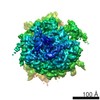
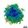
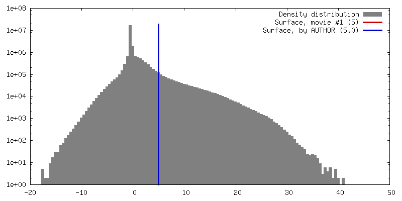
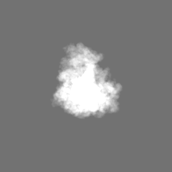
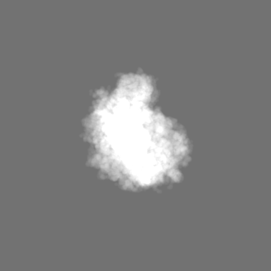
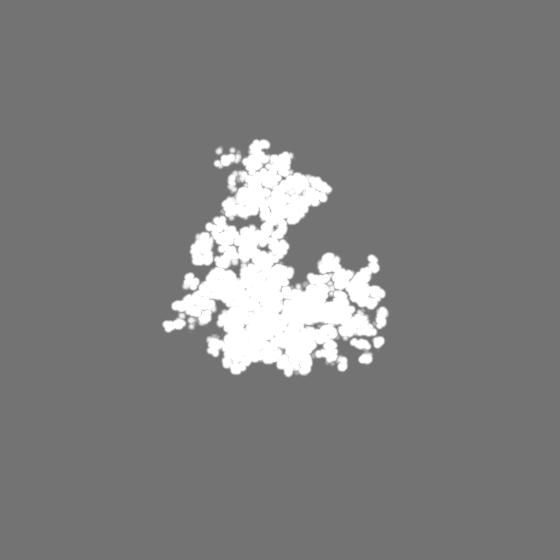
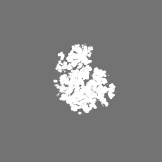
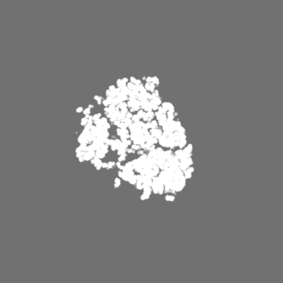
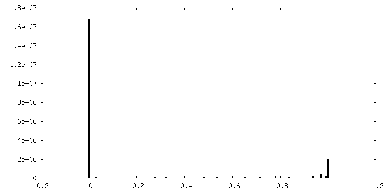
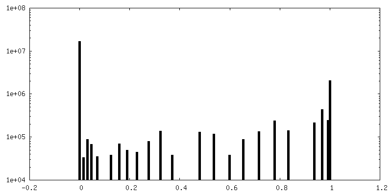
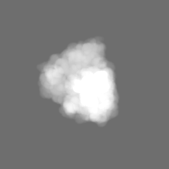
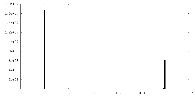
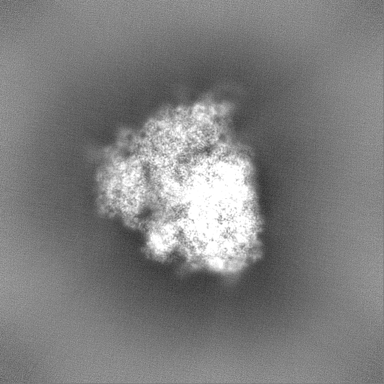
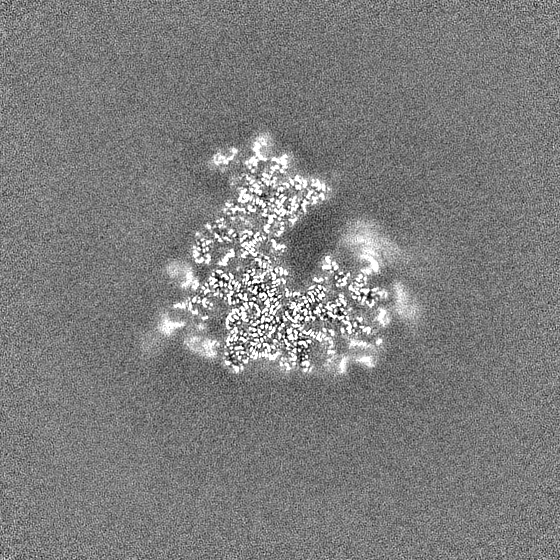
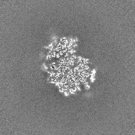
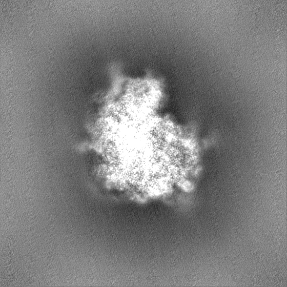
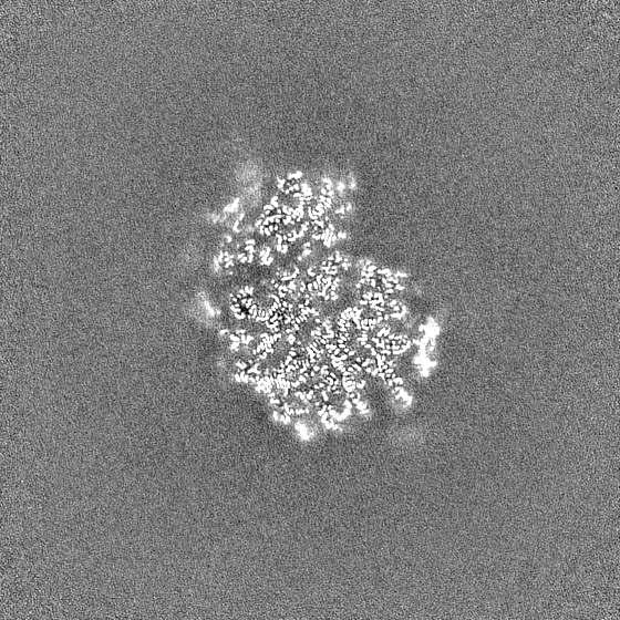
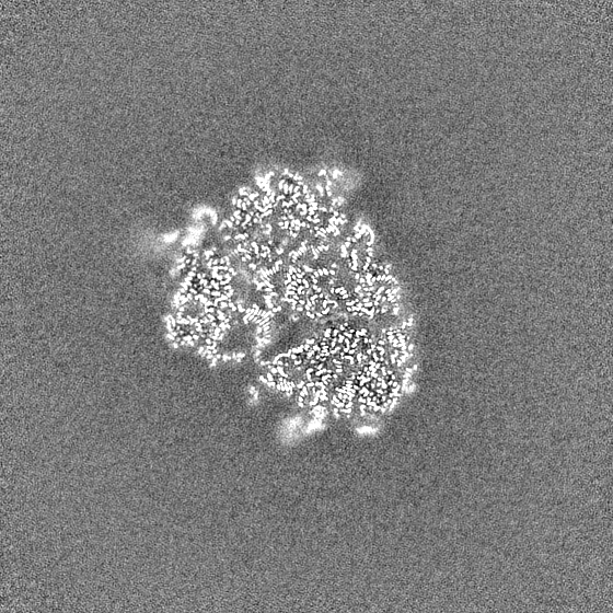
Surface view with section colored by density value
Entire : Rabbit 80S ribosome stalled close to the mutated SARS-CoV-2 slipp...
Entire
Name: Rabbit 80S ribosome stalled close to the mutated SARS-CoV-2 slippery site by a pseudoknot
Components
Complex: Rabbit 80S ribosome stalled close to the mutated SARS-CoV-2 slippery site by a pseudoknot
Complex: Rabbit 80S ribosome stalled
RNA: 18S rRNA
Protein or peptide: 40S ribosomal protein S27
Protein or peptide: Ribosomal protein S28
Protein or peptide: Ribosomal protein S27a
Protein or peptide: 40S ribosomal protein S30
Protein or peptide: Ribosomal protein eS26
Protein or peptide: RACK1
Protein or peptide: uS14
Protein or peptide: 40S ribosomal protein SA
Protein or peptide: 40S ribosomal protein S3a
Protein or peptide: Ribosomal protein uS5
Protein or peptide: 40S ribosomal protein S3
Protein or peptide: Ribosomal protein eS4
Protein or peptide: Ribosomal protein S5
Protein or peptide: 40S ribosomal protein S6
Protein or peptide: 40S ribosomal protein S7
Protein or peptide: 40S ribosomal protein S8
Protein or peptide: Ribosomal protein S9 (Predicted)
Protein or peptide: eS10
Protein or peptide: 40S ribosomal protein S11
Protein or peptide: 40S ribosomal protein S12
Protein or peptide: uS15
Protein or peptide: 40S ribosomal protein uS11
Protein or peptide: 40S ribosomal protein uS19
Protein or peptide: uS9
Protein or peptide: 40S ribosomal protein eS17
Protein or peptide: 40S ribosomal protein S18
Protein or peptide: Ribosomal protein eS19
Protein or peptide: 40S ribosomal protein uS10
Protein or peptide: Ribosomal protein eS21
Protein or peptide: Ribosomal protein S15a
Protein or peptide: 40S ribosomal protein S23
Protein or peptide: 40S ribosomal protein S24
Protein or peptide: 40S ribosomal protein S25
Protein or peptide: 60s ribosomal protein l41
RNA: 28S rRNA
RNA: 5S
RNA: 5.8S rRNA
Protein or peptide: Ribosomal protein uL2
Protein or peptide: Ribosomal protein L3
Protein or peptide: 60S ribosomal protein L4
Protein or peptide: Ribosomal_L18_c domain-containing protein
Protein or peptide: 60S ribosomal protein L6
Protein or peptide: Ribosomal Protein uL30
Protein or peptide: Ribosomal protein eL8
Protein or peptide: 60S ribosomal protein L9
Protein or peptide: 60S ribosomal protein L10
Protein or peptide: Ribosomal protein L11
Protein or peptide: Replicase polyprotein 1ab
Protein or peptide: Ribosomal protein eL13
Protein or peptide: Ribosomal protein L14
Protein or peptide: Ribosomal protein L15
Protein or peptide: Ribosomal protein uL13
Protein or peptide: uL22
Protein or peptide: Ribosomal Protein eL18
Protein or peptide: 60S ribosomal protein L19
Protein or peptide: Ribosomal protein eL20
Protein or peptide: eL21
Protein or peptide: Ribosomal protein eL22
Protein or peptide: Ribosomal protein L23
Protein or peptide: eL24
Protein or peptide: uL23
Protein or peptide: Ribosomal protein L26
Protein or peptide: 60S ribosomal protein L27
Protein or peptide: 60S ribosomal protein L27a
Protein or peptide: 60S ribosomal protein L29
Protein or peptide: eL30
Protein or peptide: eL31
Protein or peptide: eL32
Protein or peptide: eL33
Protein or peptide: 60S ribosomal protein L34
Protein or peptide: uL29
Protein or peptide: 60S ribosomal protein L36
Protein or peptide: Ribosomal protein L37
Protein or peptide: eL38
Protein or peptide: eL39
Protein or peptide: 60S ribosomal protein L40
Protein or peptide: eL42
Protein or peptide: eL43
Protein or peptide: Ribosomal protein eL28
Protein or peptide: 60S acidic ribosomal protein P0
Protein or peptide: Ribosomal protein L12
Protein or peptide: Ribosomal protein uL1
Complex: SARS-CoV-2 slippery site by a pseudoknot
RNA: mRNA containing SARS-CoV-2 sequence
Complex: Rabbit 80S ribosome stalled
RNA: E-site tRNA
RNA: P-site Phe-tRNA(Phe)
Complex: Replicase polyprotein 1ab
Ligand: SPERMIDINE
Ligand: SPERMINE
Ligand: MAGNESIUM ION
Ligand: UNKNOWN ATOM OR ION
Ligand: ZINC ION
Ligand: water
+
Supramolecule #1: Rabbit 80S ribosome stalled close to the mutated SARS-CoV-2 slipp...
Supramolecule
Name: Rabbit 80S ribosome stalled close to the mutated SARS-CoV-2 slippery site by a pseudoknot type: complex / ID: 1 / Parent: 0 / Macromolecule list: #1-#86
Model: Quantifoil R2/2 / Material: COPPER / Mesh: 400 / Support film - Material: CARBON / Support film - topology: CONTINUOUS / Support film - Film thickness: 1 / Pretreatment - Type: GLOW DISCHARGE
Vitrification
Cryogen name: ETHANE-PROPANE / Chamber humidity: 100 % / Chamber temperature: 277 K / Instrument: FEI VITROBOT MARK IV
-
Electron microscopy
Microscope
FEI TITAN KRIOS
Specialist optics
Energy filter - Name: GIF Quantum LS / Energy filter - Slit width: 20 eV
Image recording
Film or detector model: GATAN K3 (6k x 4k) / Average electron dose: 60.0 e/Å2
Electron beam
Acceleration voltage: 300 kV / Electron source: FIELD EMISSION GUN
In the structure databanks used in Yorodumi, some data are registered as the other names, "COVID-19 virus" and "2019-nCoV". Here are the details of the virus and the list of structure data.
Jan 31, 2019. EMDB accession codes are about to change! (news from PDBe EMDB page)
EMDB accession codes are about to change! (news from PDBe EMDB page)
The allocation of 4 digits for EMDB accession codes will soon come to an end. Whilst these codes will remain in use, new EMDB accession codes will include an additional digit and will expand incrementally as the available range of codes is exhausted. The current 4-digit format prefixed with “EMD-” (i.e. EMD-XXXX) will advance to a 5-digit format (i.e. EMD-XXXXX), and so on. It is currently estimated that the 4-digit codes will be depleted around Spring 2019, at which point the 5-digit format will come into force.
The EM Navigator/Yorodumi systems omit the EMD- prefix.
Related info.:Q: What is EMD? / ID/Accession-code notation in Yorodumi/EM Navigator
Yorodumi is a browser for structure data from EMDB, PDB, SASBDB, etc.
This page is also the successor to EM Navigator detail page, and also detail information page/front-end page for Omokage search.
The word "yorodu" (or yorozu) is an old Japanese word meaning "ten thousand". "mi" (miru) is to see.
Related info.:EMDB / PDB / SASBDB / Comparison of 3 databanks / Yorodumi Search / Aug 31, 2016. New EM Navigator & Yorodumi / Yorodumi Papers / Jmol/JSmol / Function and homology information / Changes in new EM Navigator and Yorodumi
 Movie
Movie Controller

 Yorodumi
 Open data
 Basic information
 Map data
 Sample
 Keywords
 Function and homology information

 Severe acute respiratory syndrome coronavirus 2 /
  Authors
 Switzerland,
  Ireland, 2 items
  Citation
 Structure visualization
 Downloads & links
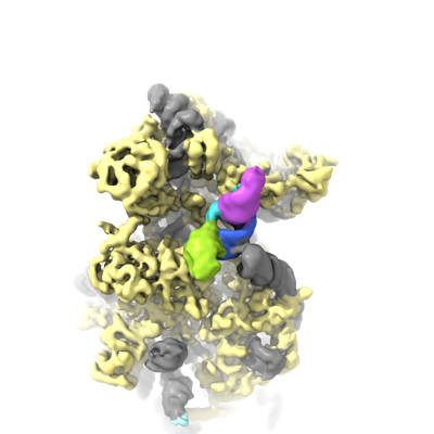 emd_12757.png
 http://ftp.pdbj.org/pub/emdb/structures/EMD-12757


 X (Sec.)
 Y (Row.)
 Z (Col.)


 Sample components


 Processing
 Electron microscopy
 FIELD EMISSION GUN