 ムービー
ムービー コントローラー


+ データを開く

- 基本情報
基本情報
| 登録情報 | データベース: EMDB / ID: EMD-12640 | |||||||||
|---|---|---|---|---|---|---|---|---|---|---|
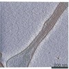
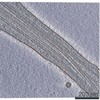
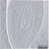
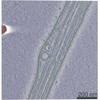
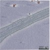
| タイトル | In situ subtomogram average of microtubule inner protein from Mus musculus DRG axons | |||||||||
 マップデータ マップデータ | microtubule inner protein structure from mouse DRG axon microtubules | |||||||||
 試料 試料 |
| |||||||||
| 生物種 |  | |||||||||
| 手法 | サブトモグラム平均法 / クライオ電子顕微鏡法 / 解像度: 32.0 Å | |||||||||
 データ登録者 データ登録者 | Foster HE / Ventura Santos C / Carter AP | |||||||||
| 資金援助 |  英国, 2件 英国, 2件
| |||||||||
 引用 引用 | ジャーナル: J Cell Biol / 年: 2022 タイトル: A cryo-ET survey of microtubules and intracellular compartments in mammalian axons. 著者: Helen E Foster / Camilla Ventura Santos / Andrew P Carter / 要旨: The neuronal axon is packed with cytoskeletal filaments, membranes, and organelles, many of which move between the cell body and axon tip. Here, we used cryo-electron tomography to survey the ...The neuronal axon is packed with cytoskeletal filaments, membranes, and organelles, many of which move between the cell body and axon tip. Here, we used cryo-electron tomography to survey the internal components of mammalian sensory axons. We determined the polarity of the axonal microtubules (MTs) by combining subtomogram classification and visual inspection, finding MT plus and minus ends are structurally similar. Subtomogram averaging of globular densities in the MT lumen suggests they have a defined structure, which is surprising given they likely contain the disordered protein MAP6. We found the endoplasmic reticulum in axons is tethered to MTs through multiple short linkers. We surveyed membrane-bound cargos and describe unexpected internal features such as granules and broken membranes. In addition, we detected proteinaceous compartments, including numerous virus-like capsid particles. Our observations outline novel features of axonal cargos and MTs, providing a platform for identification of their constituents. #1: ジャーナル: Biorxiv / 年: 2021タイトル: A cryo-ET survey of intracellular compartments within mammalian axons 著者: Foster HE / Carter AP | |||||||||
| 履歴 |
|
- 構造の表示
構造の表示
| ムービー |
 ムービービューア ムービービューア |
|---|---|
| 構造ビューア | EMマップ: SurfViewMolmilJmol/JSmol |
| 添付画像 |

- ダウンロードとリンク
-EMDBアーカイブ
| マップデータ | emd_12640.map.gz | 967.8 KB | EMDBマップデータ形式 | |
|---|---|---|---|---|
| ヘッダ (付随情報) | emd-12640-v30.xmlemd-12640.xml | 17.5 KB 17.5 KB | 表示 表示 | EMDBヘッダ |
| FSC (解像度算出) | emd_12640_fsc.xml | 2.5 KB | 表示 | FSCデータファイル |
| 画像 |  emd_12640.png emd_12640.png | 34.4 KB | ||
| マスクデータ | emd_12640_msk_1.map | 1 MB | マスクマップ | |
| その他 | emd_12640_half_map_1.map.gzemd_12640_half_map_2.map.gz | 952.9 KB 953.3 KB | ||
| アーカイブディレクトリ |  http://ftp.pdbj.org/pub/emdb/structures/EMD-12640ftp://ftp.pdbj.org/pub/emdb/structures/EMD-12640 http://ftp.pdbj.org/pub/emdb/structures/EMD-12640ftp://ftp.pdbj.org/pub/emdb/structures/EMD-12640 | HTTPS FTP |
-検証レポート
| 文書・要旨 | emd_12640_validation.pdf.gz | 415.1 KB | 表示 | EMDB検証レポート |
|---|---|---|---|---|
| 文書・詳細版 | emd_12640_full_validation.pdf.gz | 414.6 KB | 表示 | |
| XML形式データ | emd_12640_validation.xml.gz | 7.6 KB | 表示 | |
| CIF形式データ | emd_12640_validation.cif.gz | 9.4 KB | 表示 | |
| アーカイブディレクトリ | https://ftp.pdbj.org/pub/emdb/validation_reports/EMD-12640ftp://ftp.pdbj.org/pub/emdb/validation_reports/EMD-12640 | HTTPS FTP |
-関連構造データ
| 関連構造データ | C: 同じ文献を引用 ( |
|---|---|
| 類似構造データ | |
| 電子顕微鏡画像生データ | EMPIAR-10814 (タイトル: Cryo electron tomograms of mouse DRG axons (dataset 2) Data size: 206.2 Data #1: Raw image frames of mouse dorsal root ganglion axons (dataset 2) [micrographs - multiframe] Data #2: Corrected, aligned, dose-filtered and order-sorted tilt series for mouse dorsal root ganglion axons (dataset 2) [tilt series] Data #3: Tomograms of mouse dorsal root ganglion axons (dataset 2) binned by 4 and deconvolved [reconstructed volumes]) |


-リンク
| EMDBのページ | EMDB (EBI/PDBe) / EMDataResource |
|---|
-マップ
| ファイル | ダウンロード / ファイル: emd_12640.map.gz / 形式: CCP4 / 大きさ: 1 MB / タイプ: IMAGE STORED AS FLOATING POINT NUMBER (4 BYTES) | ||||||||||||||||||||||||||||||||||||||||||||||||||||||||||||
|---|---|---|---|---|---|---|---|---|---|---|---|---|---|---|---|---|---|---|---|---|---|---|---|---|---|---|---|---|---|---|---|---|---|---|---|---|---|---|---|---|---|---|---|---|---|---|---|---|---|---|---|---|---|---|---|---|---|---|---|---|---|
| 注釈 | microtubule inner protein structure from mouse DRG axon microtubules | ||||||||||||||||||||||||||||||||||||||||||||||||||||||||||||
| 投影像・断面図 | 画像のコントロール
画像は Spider により作成 | ||||||||||||||||||||||||||||||||||||||||||||||||||||||||||||
| ボクセルのサイズ | X=Y=Z: 5.5 Å | ||||||||||||||||||||||||||||||||||||||||||||||||||||||||||||
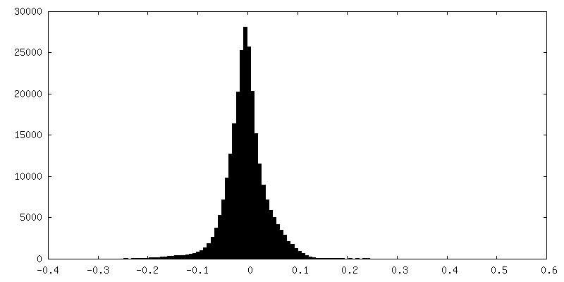
| 密度 |
| ||||||||||||||||||||||||||||||||||||||||||||||||||||||||||||
| 対称性 | 空間群: 1 | ||||||||||||||||||||||||||||||||||||||||||||||||||||||||||||
| 詳細 | EMDB XML:
CCP4マップ ヘッダ情報:
| ||||||||||||||||||||||||||||||||||||||||||||||||||||||||||||
 Z (Sec.)
 Y (Row.)
 X (Col.)


-添付データ
-マスク #1
| ファイル | emd_12640_msk_1.map | ||||||||||||
|---|---|---|---|---|---|---|---|---|---|---|---|---|---|
| 投影像・断面図 |
| ||||||||||||
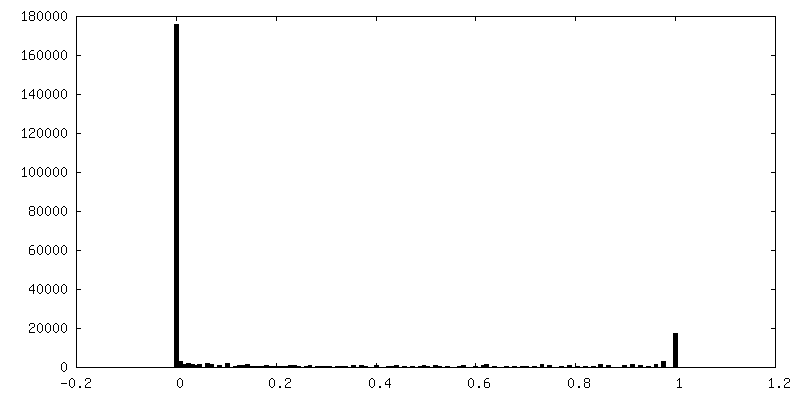
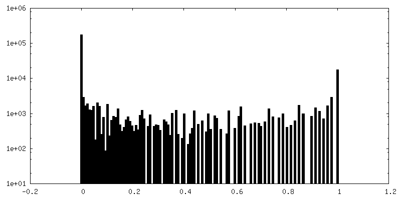
| 密度ヒストグラム |


-ハーフマップ: half map1
| ファイル | emd_12640_half_map_1.map | ||||||||||||
|---|---|---|---|---|---|---|---|---|---|---|---|---|---|
| 注釈 | half map1 | ||||||||||||
| 投影像・断面図 |
| ||||||||||||
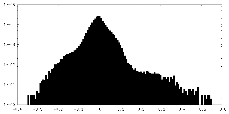
| 密度ヒストグラム |


-ハーフマップ: half map2
| ファイル | emd_12640_half_map_2.map | ||||||||||||
|---|---|---|---|---|---|---|---|---|---|---|---|---|---|
| 注釈 | half map2 | ||||||||||||
| 投影像・断面図 |
| ||||||||||||
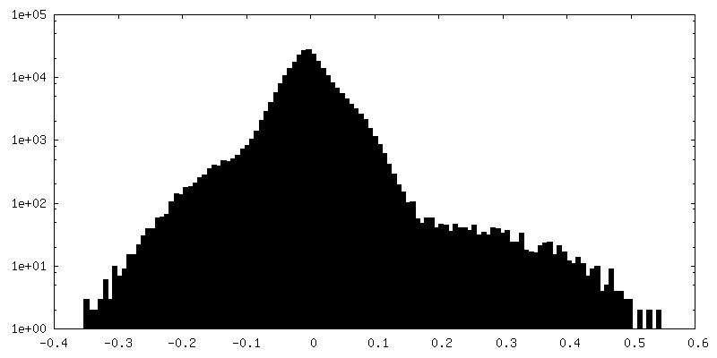
| 密度ヒストグラム |


- 試料の構成要素
-全体 : In situ subtomogram average of microtubule inner protein from Mus...
| 全体 | 名称: In situ subtomogram average of microtubule inner protein from Mus musculus DRG axons |
|---|---|
| 要素 |
|
-超分子 #1: In situ subtomogram average of microtubule inner protein from Mus...
| 超分子 | 名称: In situ subtomogram average of microtubule inner protein from Mus musculus DRG axons タイプ: organelle_or_cellular_component / ID: 1 / 親要素: 0 / 含まれる分子: #1 |
|---|---|
| 由来(天然) | 生物種: |
-実験情報
-構造解析
| 手法 | クライオ電子顕微鏡法 |
|---|---|
 解析 解析 | サブトモグラム平均法 |
| 試料の集合状態 | cell |
-試料調製
| 緩衝液 | pH: 7.4 |
|---|---|
| グリッド | モデル: Quantifoil R3.5/1 / 材質: GOLD / メッシュ: 200 / 支持フィルム - 材質: CARBON / 支持フィルム - トポロジー: CONTINUOUS / 前処理 - タイプ: PLASMA CLEANING / 前処理 - 雰囲気: OTHER 詳細: Grids were additionally coated in 0.1mg/mL poly-L-lysine then 0.01mg/mL laminin before plating |
| 凍結 | 凍結剤: ETHANE / チャンバー内湿度: 100 % / チャンバー内温度: 310 K / 装置: FEI VITROBOT MARK IV / 詳細: Manual blot for 3 s before plunging. |
| 詳細 | Microtubule inner proteins in axons of adult DRG neurons grown for 7 days in vitro |
- 電子顕微鏡法
| 顕微鏡 | FEI TITAN KRIOS |
|---|---|
| 特殊光学系 | エネルギーフィルター - 名称: GIF Bioquantum / エネルギーフィルター - スリット幅: 20 eV |
| 撮影 | フィルム・検出器のモデル: GATAN K2 SUMMIT (4k x 4k) 検出モード: COUNTING / デジタル化 - サイズ - 横: 3710 pixel / デジタル化 - サイズ - 縦: 3838 pixel / デジタル化 - 画像ごとのフレーム数: 1-10 / 平均露光時間: 1.7 sec. / 平均電子線量: 1.85 e/Å2 詳細: 61 images per tilt series with 112.85 e/A2 total dose. |
| 電子線 | 加速電圧: 300 kV / 電子線源:  FIELD EMISSION GUN FIELD EMISSION GUN |
| 電子光学系 | C2レンズ絞り径: 50.0 µm / 照射モード: FLOOD BEAM / 撮影モード: BRIGHT FIELD / Cs: 2.7 mm / 最大 デフォーカス(公称値): 5.0 µm / 最小 デフォーカス(公称値): 3.0 µm / 倍率(公称値): 53000 |
| 試料ステージ | 試料ホルダーモデル: FEI TITAN KRIOS AUTOGRID HOLDER ホルダー冷却材: NITROGEN |
| 実験機器 |  モデル: Titan Krios / 画像提供: FEI Company |

