- EMDB-12455: The cryoEM density map hexamer from PFO perforated VLPs in the pr... -
+
Open data
ID or keywords:
Loading...
-
Basic information
Entry
Database: EMDB / ID: EMD-12455
Title
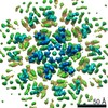
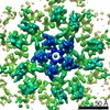
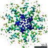
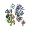
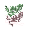
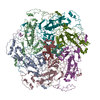
The cryoEM density map hexamer from PFO perforated VLPs in the presence of IP6/CypA-DsRed
Map data
half1 and half2 merged map low pass to 6 angstrom and b-factor -100
Sample
Virus: Human immunodeficiency virus 1
Protein or peptide: HIV-1 Capsid
Function / homology
Function and homology information
integrase activity / Integration of viral DNA into host genomic DNA / Autointegration results in viral DNA circles / Minus-strand DNA synthesis / Plus-strand DNA synthesis / Uncoating of the HIV Virion / 2-LTR circle formation / Vpr-mediated nuclear import of PICs / Early Phase of HIV Life Cycle / Integration of provirus ...integrase activity / Integration of viral DNA into host genomic DNA / Autointegration results in viral DNA circles / Minus-strand DNA synthesis / Plus-strand DNA synthesis / Uncoating of the HIV Virion / 2-LTR circle formation / Vpr-mediated nuclear import of PICs / Early Phase of HIV Life Cycle / Integration of provirus / APOBEC3G mediated resistance to HIV-1 infection / Binding and entry of HIV virion / viral life cycle / HIV-1 retropepsin / symbiont-mediated activation of host apoptosis / retroviral ribonuclease H / exoribonuclease H / exoribonuclease H activity / Assembly Of The HIV Virion / protein processing / viral genome integration into host DNA / Budding and maturation of HIV virion / establishment of integrated proviral latency / RNA-directed DNA polymerase / RNA stem-loop binding / viral penetration into host nucleus / host multivesicular body / RNA-directed DNA polymerase activity / RNA-DNA hybrid ribonuclease activity / Transferases; Transferring phosphorus-containing groups; Nucleotidyltransferases / peptidase activity / host cell / viral nucleocapsid / DNA recombination / DNA-directed DNA polymerase / aspartic-type endopeptidase activity / Hydrolases; Acting on ester bonds / host cell cytoplasm / DNA-directed DNA polymerase activity / symbiont-mediated suppression of host gene expression / viral translational frameshifting / symbiont entry into host cell / lipid binding / host cell nucleus / host cell plasma membrane / virion membrane / structural molecule activity / DNA binding / RNA binding / zinc ion binding / identical protein binding Similarity search - Function
Biotechnology and Biological Sciences Research Council (BBSRC)
BB/S003339/1
United Kingdom
Wellcome Trust
206422/Z/17/Z
United Kingdom
Wellcome Trust
203141/Z/16/Z
United Kingdom
Citation
Journal: Sci Adv / Year: 2021 Title: Structure of native HIV-1 cores and their interactions with IP6 and CypA. Authors: Tao Ni / Yanan Zhu / Zhengyi Yang / Chaoyi Xu / Yuriy Chaban / Tanya Nesterova / Jiying Ning / Till Böcking / Michael W Parker / Christina Monnie / Jinwoo Ahn / Juan R Perilla / Peijun Zhang / Abstract: The viral capsid plays essential roles in HIV replication and is a major platform engaging host factors. To overcome challenges in study native capsid structure, we used the perfringolysin O to ...The viral capsid plays essential roles in HIV replication and is a major platform engaging host factors. To overcome challenges in study native capsid structure, we used the perfringolysin O to perforate the membrane of HIV-1 particles, thus allowing host proteins and small molecules to access the native capsid while improving cryo–electron microscopy image quality. Using cryo–electron tomography and subtomogram averaging, we determined the structures of native capsomers in the presence and absence of inositol hexakisphosphate (IP6) and cyclophilin A and constructed an all-atom model of a complete HIV-1 capsid. Our structures reveal two IP6 binding sites and modes of cyclophilin A interactions. Free energy calculations substantiate the two binding sites at R18 and K25 and further show a prohibitive energy barrier for IP6 to pass through the pentamer. Our results demonstrate that perfringolysin O perforation is a valuable tool for structural analyses of enveloped virus capsids and interactions with host cell factors.
History
Deposition
Feb 20, 2021
-
Header (metadata) release
Jun 2, 2021
-
Map release
Jun 2, 2021
-
Update
Dec 1, 2021
-
Current status
Dec 1, 2021
Processing site: PDBe / Status: Released
-
Structure visualization
Movie
Surface view with section colored by density value
In the structure databanks used in Yorodumi, some data are registered as the other names, "COVID-19 virus" and "2019-nCoV". Here are the details of the virus and the list of structure data.
Jan 31, 2019. EMDB accession codes are about to change! (news from PDBe EMDB page)
EMDB accession codes are about to change! (news from PDBe EMDB page)
The allocation of 4 digits for EMDB accession codes will soon come to an end. Whilst these codes will remain in use, new EMDB accession codes will include an additional digit and will expand incrementally as the available range of codes is exhausted. The current 4-digit format prefixed with “EMD-” (i.e. EMD-XXXX) will advance to a 5-digit format (i.e. EMD-XXXXX), and so on. It is currently estimated that the 4-digit codes will be depleted around Spring 2019, at which point the 5-digit format will come into force.
The EM Navigator/Yorodumi systems omit the EMD- prefix.
Related info.:Q: What is EMD? / ID/Accession-code notation in Yorodumi/EM Navigator
Yorodumi is a browser for structure data from EMDB, PDB, SASBDB, etc.
This page is also the successor to EM Navigator detail page, and also detail information page/front-end page for Omokage search.
The word "yorodu" (or yorozu) is an old Japanese word meaning "ten thousand". "mi" (miru) is to see.
Related info.:EMDB / PDB / SASBDB / Comparison of 3 databanks / Yorodumi Search / Aug 31, 2016. New EM Navigator & Yorodumi / Yorodumi Papers / Jmol/JSmol / Function and homology information / Changes in new EM Navigator and Yorodumi
 Movie
Movie Controller

 Yorodumi
 Open data
 Basic information
 Map data
 Sample
 Function and homology information
 Human immunodeficiency virus 1
 Authors
 United Kingdom, 3 items
  Citation

 Structure visualization
 Downloads & links
 emd_12455.png
 http://ftp.pdbj.org/pub/emdb/structures/EMD-12455


 Z (Sec.)
 Y (Row.)
 X (Col.)


 Sample components
 Homo sapiens (human)
 Processing
 Electron microscopy
 FIELD EMISSION GUN
