Movie
Movie Controller

[English] 日本語
 Yorodumi
- EMDB-1213: Structural model for the mannose receptor family uncovered by ele... -
+ Open data

- Basic information
| Entry | Database: EMDB / ID: EMD-1213 | |||||||||
|---|---|---|---|---|---|---|---|---|---|---|
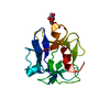
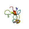
| Title | Structural model for the mannose receptor family uncovered by electron microscopy of Endo180 and the mannose receptor. | |||||||||
 Map data Map data | This a 3D reconstruction of the soluble fragment of the mannose receptor | |||||||||
 Sample Sample |
| |||||||||
| Biological species |  | |||||||||
| Method | single particle reconstruction / negative staining / Resolution: 33.0 Å | |||||||||
 Authors Authors | Boskovic J / Arnold JN / Stilion R / Gordon S / Sim RB / Rivera-Calzada A / Wienke D / Isacke CM / Martinez-Pomares L / Llorca O | |||||||||
 Citation Citation | Journal: J Biol Chem / Year: 2006 Title: Structural model for the mannose receptor family uncovered by electron microscopy of Endo180 and the mannose receptor. Authors: Jasminka Boskovic / James N Arnold / Richard Stilion / Siamon Gordon / Robert B Sim / Angel Rivera-Calzada / Dirk Wienke / Clare M Isacke / Luisa Martinez-Pomares / Oscar Llorca /  Abstract: The mannose receptor family comprises four members in mammals, Endo180 (CD280), DEC-205 (CD205), phospholipase A(2) receptor (PLA(2)R) and the mannose receptor (MR, CD206), whose extracellular ...The mannose receptor family comprises four members in mammals, Endo180 (CD280), DEC-205 (CD205), phospholipase A(2) receptor (PLA(2)R) and the mannose receptor (MR, CD206), whose extracellular portion contains a similar domain arrangement: an N-terminal cysteine-rich domain (CysR) followed by a single fibronectin type II domain (FNII) and 8-10 C-type lectin-like domains (CTLDs). These proteins mediate diverse functions ranging from extracellular matrix turnover through collagen uptake to homeostasis and immunity based on sugar recognition. Endo180 and the MR are multivalent transmembrane receptors capable of interacting with multiple ligands; in both receptors FNII recognizes collagens, and a single CTLD retains lectin activity (CTLD2 in Endo180 and CTLD4 in MR). It is expected that the overall conformation of these multivalent molecules would deeply influence their function as the availability of their binding sites could be altered under different conditions. However, conflicting reports have been published on the three-dimensional arrangement of these receptors. Here, we have used single particle electron microscopy to elucidate the three-dimensional organization of the MR and Endo180. Strikingly, we have found that both receptors display distinct three-dimensional structures, which are, however, conceptually very similar: a bent and compact conformation built upon interactions of the CysR domain and the lone functional CTLD. Biochemical and electron microscopy experiments indicate that, under a low pH mimicking the endosomal environment, both MR and Endo180 experience large conformational changes. We propose a structural model for the mannose receptor family where at least two conformations exist that may serve to regulate differences in ligand selectivity. | |||||||||
| History |
|
- Structure visualization
| Movie |
 Movie viewer Movie viewer |
|---|---|
| Structure viewer | EM map: SurfViewMolmilJmol/JSmol |
| Supplemental images |
 UCSF Chimera
- Downloads & links
-EMDB archive
| Map data | emd_1213.map.gz | 216.2 KB | EMDB map data format | |
|---|---|---|---|---|
| Header (meta data) | emd-1213-v30.xmlemd-1213.xml | 10.7 KB 10.7 KB | Display Display | EMDB header |
| Images |  1213.gif 1213.gif | 39.1 KB | ||
| Archive directory |  http://ftp.pdbj.org/pub/emdb/structures/EMD-1213ftp://ftp.pdbj.org/pub/emdb/structures/EMD-1213 http://ftp.pdbj.org/pub/emdb/structures/EMD-1213ftp://ftp.pdbj.org/pub/emdb/structures/EMD-1213 | HTTPS FTP |
-Related structure data
| Similar structure data |
|---|


-Links
| EMDB pages | EMDB (EBI/PDBe) / EMDataResource |
|---|
-Map
| File | Download / File: emd_1213.map.gz / Format: CCP4 / Size: 422.9 KB / Type: IMAGE STORED AS FLOATING POINT NUMBER (4 BYTES) | ||||||||||||||||||||||||||||||||||||||||||||||||||||||||||||||||||||
|---|---|---|---|---|---|---|---|---|---|---|---|---|---|---|---|---|---|---|---|---|---|---|---|---|---|---|---|---|---|---|---|---|---|---|---|---|---|---|---|---|---|---|---|---|---|---|---|---|---|---|---|---|---|---|---|---|---|---|---|---|---|---|---|---|---|---|---|---|---|
| Annotation | This a 3D reconstruction of the soluble fragment of the mannose receptor | ||||||||||||||||||||||||||||||||||||||||||||||||||||||||||||||||||||
| Projections & slices | Image control
Images are generated by Spider. | ||||||||||||||||||||||||||||||||||||||||||||||||||||||||||||||||||||
| Voxel size | X=Y=Z: 5.3 Å | ||||||||||||||||||||||||||||||||||||||||||||||||||||||||||||||||||||
| Density |
| ||||||||||||||||||||||||||||||||||||||||||||||||||||||||||||||||||||
| Symmetry | Space group: 1 | ||||||||||||||||||||||||||||||||||||||||||||||||||||||||||||||||||||
| Details | EMDB XML:
CCP4 map header:
| ||||||||||||||||||||||||||||||||||||||||||||||||||||||||||||||||||||
 Z (Sec.)
 Y (Row.)
 X (Col.)


-Supplemental data
- Sample components
-Entire : Soluble Form of Mouse Mannose Receptor
| Entire | Name: Soluble Form of Mouse Mannose Receptor |
|---|---|
| Components |
|
-Supramolecule #1000: Soluble Form of Mouse Mannose Receptor
| Supramolecule | Name: Soluble Form of Mouse Mannose Receptor / type: sample / ID: 1000 / Oligomeric state: monomer / Number unique components: 1 |
|---|---|
| Molecular weight | Experimental: 160 KDa / Theoretical: 180 KDa / Method: Gel Filtration Chromatography |
-Macromolecule #1: Mannose Receptor
| Macromolecule | Name: Mannose Receptor / type: protein_or_peptide / ID: 1 / Name.synonym: MR Details: MR(CD206) is a member of mannose receptor family of transmembrane receptors which acts as a molecular scavenger that binds and internalises potentially harmful glycoproteins and may also ...Details: MR(CD206) is a member of mannose receptor family of transmembrane receptors which acts as a molecular scavenger that binds and internalises potentially harmful glycoproteins and may also mediate phagocytosis of a wide variety of microbes. Number of copies: 1 / Oligomeric state: Monomer / Recombinant expression: Yes |
|---|---|
| Source (natural) | Organism: |
| Molecular weight | Experimental: 180 KDa / Theoretical: 160 KDa |
| Recombinant expression | Organism: 293T / Recombinant plasmid: pMH |
-Experimental details
-Structure determination
| Method | negative staining |
|---|---|
 Processing Processing | single particle reconstruction |
| Aggregation state | particle |
-Sample preparation
| Concentration | 0.2 mg/mL |
|---|---|
| Buffer | pH: 7.4 / Details: 10mMTris,140mM Nacl,10 mM CaCl2 |
| Staining | Type: NEGATIVE Details: Protein was adsorbed to glow-discharged carbon-coated grids and negatively stained with 2% w/v uranyl acetate |
| Grid | Details: 400 mesh copper-rhodium grid |
| Vitrification | Cryogen name: NONE |
- Electron microscopy
| Microscope | JEOL 1230 |
|---|---|
| Alignment procedure | Legacy - Astigmatism: objective lens astigmatism was corrected at 100,000 times magnification |
| Details | Electron microscope was JEOL JEM-120, 120kV. Specimen holder, JEOL type M: 207EM-11020. Images were colected in low-dose conditions |
| Image recording | Category: FILM / Film or detector model: KODAK 4489 FILM / Digitization - Scanner: OTHER / Digitization - Sampling interval: 5.3 µm / Details: Minolta Dimage Scan Multi Pro. Scan at 2400 dpi / Bits/pixel: 16 |
| Electron beam | Acceleration voltage: 100 kV / Electron source: TUNGSTEN HAIRPIN |
| Electron optics | Calibrated magnification: 38000 / Illumination mode: FLOOD BEAM / Imaging mode: BRIGHT FIELD / Cs: 2.9 mm / Nominal magnification: 40000 |
| Sample stage | Specimen holder: Eucentric / Specimen holder model: OTHER |
-Image processing
| Details | Particles were selected using boxer program from EMAN (J.Struct.Biol. (1999) 128:82-97) |
|---|---|
| Final reconstruction | Applied symmetry - Point group: C1 (asymmetric) / Algorithm: OTHER / Resolution.type: BY AUTHOR / Resolution: 33.0 Å / Resolution method: FSC 0.5 CUT-OFF / Software - Name: EMAN Details: Angular refinement using a blob as starting reference volume Number images used: 7322 |