 Movie
Movie Controller

[English] 日本語
 Yorodumi
- EMDB-12067: triplet microtubule from the proximal region of the pig sperm pro... -
+ Open data

- Basic information
| Entry | Database: EMDB / ID: EMD-12067 | |||||||||
|---|---|---|---|---|---|---|---|---|---|---|
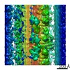
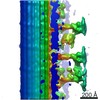
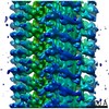
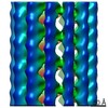
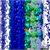
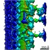
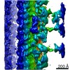
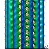
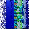
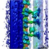
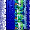
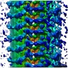
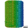
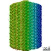
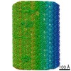
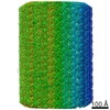
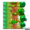
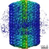
| Title | triplet microtubule from the proximal region of the pig sperm proximal centriole | |||||||||
 Map data Map data | subtomogram average of triplet microtubules from the proximal region of the pig sperm proximal centriole | |||||||||
 Sample Sample |
| |||||||||
| Biological species |  | |||||||||
| Method | subtomogram averaging / cryo EM / Resolution: 30.0 Å | |||||||||
 Authors Authors | Leung MR / Zeev-Ben-Mordehai T | |||||||||
| Funding support |  Netherlands, 1 items Netherlands, 1 items
| |||||||||
 Citation Citation | Journal: EMBO J / Year: 2021 Title: The multi-scale architecture of mammalian sperm flagella and implications for ciliary motility. Authors: Miguel Ricardo Leung / Marc C Roelofs / Ravi Teja Ravi / Paula Maitan / Heiko Henning / Min Zhang / Elizabeth G Bromfield / Stuart C Howes / Bart M Gadella / Hermes Bloomfield-Gadêlha / ...Authors: Miguel Ricardo Leung / Marc C Roelofs / Ravi Teja Ravi / Paula Maitan / Heiko Henning / Min Zhang / Elizabeth G Bromfield / Stuart C Howes / Bart M Gadella / Hermes Bloomfield-Gadêlha / Tzviya Zeev-Ben-Mordehai /    Abstract: Motile cilia are molecular machines used by a myriad of eukaryotic cells to swim through fluid environments. However, available molecular structures represent only a handful of cell types, limiting ...Motile cilia are molecular machines used by a myriad of eukaryotic cells to swim through fluid environments. However, available molecular structures represent only a handful of cell types, limiting our understanding of how cilia are modified to support motility in diverse media. Here, we use cryo-focused ion beam milling-enabled cryo-electron tomography to image sperm flagella from three mammalian species. We resolve in-cell structures of centrioles, axonemal doublets, central pair apparatus, and endpiece singlets, revealing novel protofilament-bridging microtubule inner proteins throughout the flagellum. We present native structures of the flagellar base, which is crucial for shaping the flagellar beat. We show that outer dense fibers are directly coupled to microtubule doublets in the principal piece but not in the midpiece. Thus, mammalian sperm flagella are ornamented across scales, from protofilament-bracing structures reinforcing microtubules at the nano-scale to accessory structures that impose micron-scale asymmetries on the entire assembly. Our structures provide vital foundations for linking molecular structure to ciliary motility and evolution. #1: Journal: Biorxiv / Year: 2020Title: The multi-scale architecture of mammalian sperm flagella and implications for ciliary motility Authors: Leung MR / Roelofs MC / Ravi RT / Maitan P / Zhang M / Henning H / Bromfield EG / Howes SC / Gadella BM / Bloomfield-Gadelha H / Zeev-Ben-Mordehai T | |||||||||
| History |
|
- Structure visualization
| Movie |
 Movie viewer Movie viewer |
|---|---|
| Structure viewer | EM map: SurfViewMolmilJmol/JSmol |
| Supplemental images |

- Downloads & links
-EMDB archive
| Map data | emd_12067.map.gz | 1.1 MB | EMDB map data format | |
|---|---|---|---|---|
| Header (meta data) | emd-12067-v30.xmlemd-12067.xml | 10.6 KB 10.6 KB | Display Display | EMDB header |
| Images | 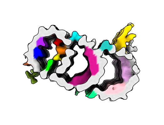 emd_12067.png emd_12067.png | 109.4 KB | ||
| Archive directory |  http://ftp.pdbj.org/pub/emdb/structures/EMD-12067ftp://ftp.pdbj.org/pub/emdb/structures/EMD-12067 http://ftp.pdbj.org/pub/emdb/structures/EMD-12067ftp://ftp.pdbj.org/pub/emdb/structures/EMD-12067 | HTTPS FTP |
-Validation report
| Summary document | emd_12067_validation.pdf.gz | 228.5 KB | Display | EMDB validaton report |
|---|---|---|---|---|
| Full document | emd_12067_full_validation.pdf.gz | 227.6 KB | Display | |
| Data in XML | emd_12067_validation.xml.gz | 4.7 KB | Display | |
| Arichive directory | https://ftp.pdbj.org/pub/emdb/validation_reports/EMD-12067ftp://ftp.pdbj.org/pub/emdb/validation_reports/EMD-12067 | HTTPS FTP |
-Related structure data
| Related structure data | C: citing same article ( |
|---|---|
| Similar structure data |
-Links
| EMDB pages | EMDB (EBI/PDBe) / EMDataResource |
|---|
-Map
| File | Download / File: emd_12067.map.gz / Format: CCP4 / Size: 1.2 MB / Type: IMAGE STORED AS FLOATING POINT NUMBER (4 BYTES) | ||||||||||||||||||||||||||||||||||||||||||||||||||||||||||||
|---|---|---|---|---|---|---|---|---|---|---|---|---|---|---|---|---|---|---|---|---|---|---|---|---|---|---|---|---|---|---|---|---|---|---|---|---|---|---|---|---|---|---|---|---|---|---|---|---|---|---|---|---|---|---|---|---|---|---|---|---|---|
| Annotation | subtomogram average of triplet microtubules from the proximal region of the pig sperm proximal centriole | ||||||||||||||||||||||||||||||||||||||||||||||||||||||||||||
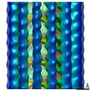
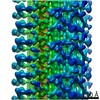
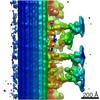
| Projections & slices | Image control
Images are generated by Spider. generated in cubic-lattice coordinate | ||||||||||||||||||||||||||||||||||||||||||||||||||||||||||||
| Voxel size | X=Y=Z: 8.68 Å | ||||||||||||||||||||||||||||||||||||||||||||||||||||||||||||
| Density |
| ||||||||||||||||||||||||||||||||||||||||||||||||||||||||||||
| Symmetry | Space group: 1 | ||||||||||||||||||||||||||||||||||||||||||||||||||||||||||||
| Details | EMDB XML:
CCP4 map header:
| ||||||||||||||||||||||||||||||||||||||||||||||||||||||||||||
 Z (Sec.)
 Y (Row.)
 X (Col.)


-Supplemental data
- Sample components
-Entire : triplet microtubules from the proximal region of the pig sperm pr...
| Entire | Name: triplet microtubules from the proximal region of the pig sperm proximal centriole |
|---|---|
| Components |
|
-Supramolecule #1: triplet microtubules from the proximal region of the pig sperm pr...
| Supramolecule | Name: triplet microtubules from the proximal region of the pig sperm proximal centriole type: organelle_or_cellular_component / ID: 1 / Parent: 0 |
|---|---|
| Source (natural) | Organism: |
-Experimental details
-Structure determination
| Method | cryo EM |
|---|---|
 Processing Processing | subtomogram averaging |
| Aggregation state | cell |
-Sample preparation
| Buffer | pH: 7.4 |
|---|---|
| Grid | Model: Quantifoil |
| Vitrification | Cryogen name: ETHANE-PROPANE / Instrument: HOMEMADE PLUNGER |
- Electron microscopy
| Microscope | FEI TALOS ARCTICA |
|---|---|
| Image recording | Film or detector model: GATAN K2 SUMMIT (4k x 4k) / Detector mode: COUNTING / Average electron dose: 1.5 e/Å2 |
| Electron beam | Acceleration voltage: 200 kV / Electron source:  FIELD EMISSION GUN FIELD EMISSION GUN |
| Electron optics | Illumination mode: FLOOD BEAM / Imaging mode: BRIGHT FIELD |
| Sample stage | Cooling holder cryogen: NITROGEN |
| Experimental equipment |  Model: Talos Arctica / Image courtesy: FEI Company |
-Image processing
| Final reconstruction | Applied symmetry - Point group: C1 (asymmetric) / Resolution.type: BY AUTHOR / Resolution: 30.0 Å / Resolution method: FSC 0.5 CUT-OFF / Number subtomograms used: 200 |
|---|---|
| Extraction | Number tomograms: 3 / Number images used: 200 / Software - Name: PEET (ver. 1.13.0) |
| CTF correction | Software - Name: PEET (ver. 1.13.0) |
| Final angle assignment | Type: OTHER / Software - Name: PEET (ver. 1.13.0) |
