[English] 日本語
Yorodumi
- EMDB-1201: Three-dimensional structure of the myosin V inhibited state by cr... -

+
Open data


ID or keywords:

Loading...

-
Basic information

Entry
Database: EMDB / ID: EMD-1201
TitleThree-dimensional structure of the myosin V inhibited state by cryoelectron tomography.
Map dataThis the 3-D average map of myosin-V in "off" state
Sample
  • Sample: MYOSIN V
  • Protein or peptide: Myosin-V
Function / homology
Function and homology information


CaMK IV-mediated phosphorylation of CREB / Cam-PDE 1 activation / CREB1 phosphorylation through the activation of CaMKII/CaMKK/CaMKIV cascasde / Glycogen breakdown (glycogenolysis) / Activation of RAC1 downstream of NMDARs / Reduction of cytosolic Ca++ levels / Sodium/Calcium exchangers / Activation of Ca-permeable Kainate Receptor / CLEC7A (Dectin-1) induces NFAT activation / Synthesis of IP3 and IP4 in the cytosol ...CaMK IV-mediated phosphorylation of CREB / Cam-PDE 1 activation / CREB1 phosphorylation through the activation of CaMKII/CaMKK/CaMKIV cascasde / Glycogen breakdown (glycogenolysis) / Activation of RAC1 downstream of NMDARs / Reduction of cytosolic Ca++ levels / Sodium/Calcium exchangers / Activation of Ca-permeable Kainate Receptor / CLEC7A (Dectin-1) induces NFAT activation / Synthesis of IP3 and IP4 in the cytosol / RHO GTPases activate PAKs / Calmodulin induced events / Inactivation, recovery and regulation of the phototransduction cascade / Tetrahydrobiopterin (BH4) synthesis, recycling, salvage and regulation / Calcineurin activates NFAT / eNOS activation / Ion transport by P-type ATPases / Unblocking of NMDA receptors, glutamate binding and activation / Protein methylation / RAF activation / VEGFR2 mediated vascular permeability / RAS processing / Smooth Muscle Contraction / minus-end directed microfilament motor activity / Ca2+ pathway / negative regulation of calcium ion transmembrane transporter activity / FCERI mediated Ca+2 mobilization / RAF/MAP kinase cascade / RHO GTPases activate IQGAPs / Extra-nuclear estrogen signaling / insulin-responsive compartment / PKA activation / regulation of response to tumor cell / positive regulation of autophagic cell death / DAPK1-calmodulin complex / Platelet degranulation / : / establishment of protein localization to mitochondrial membrane / Stimuli-sensing channels / Ion homeostasis / type 3 metabotropic glutamate receptor binding / vesicle transport along actin filament / myosin complex / regulation of synaptic vesicle endocytosis / microfilament motor activity / negative regulation of high voltage-gated calcium channel activity / regulation of synaptic vesicle exocytosis / negative regulation of calcium ion export across plasma membrane / organelle localization by membrane tethering / regulation of cardiac muscle cell action potential / mitochondrion-endoplasmic reticulum membrane tethering / autophagosome membrane docking / response to corticosterone / positive regulation of ryanodine-sensitive calcium-release channel activity / nitric-oxide synthase binding / filamentous actin / protein phosphatase activator activity / positive regulation of cyclic-nucleotide phosphodiesterase activity / positive regulation of phosphoprotein phosphatase activity / adenylate cyclase binding / catalytic complex / detection of calcium ion / negative regulation of ryanodine-sensitive calcium-release channel activity / regulation of cardiac muscle contraction / calcium channel inhibitor activity / cellular response to interferon-beta / enzyme regulator activity / voltage-gated potassium channel complex / phosphatidylinositol 3-kinase binding / regulation of release of sequestered calcium ion into cytosol by sarcoplasmic reticulum / regulation of calcium-mediated signaling / positive regulation of protein dephosphorylation / titin binding / regulation of ryanodine-sensitive calcium-release channel activity / vesicle-mediated transport / potassium ion transmembrane transport / sperm midpiece / response to amphetamine / calcium channel complex / activation of adenylate cyclase activity / adenylate cyclase activator activity / regulation of heart rate / nitric-oxide synthase regulator activity / protein serine/threonine kinase activator activity / sarcomere / regulation of cytokinesis / actin filament organization / positive regulation of nitric-oxide synthase activity / calcium-mediated signaling / spindle microtubule / protein localization to plasma membrane / mitochondrial membrane / positive regulation of receptor signaling pathway via JAK-STAT / synaptic vesicle membrane / spindle pole / cellular response to type II interferon / response to calcium ion / cellular response to insulin stimulus / calcium-dependent protein binding / disordered domain specific binding
Similarity search - Function
Myosin 5a, cargo-binding domain / Class V myosin, motor domain / Dilute domain / DIL domain / Dilute domain profile. / DIL / IQ calmodulin-binding motif / Myosin, N-terminal, SH3-like / Myosin N-terminal SH3-like domain profile. / Short calmodulin-binding motif containing conserved Ile and Gln residues. ...Myosin 5a, cargo-binding domain / Class V myosin, motor domain / Dilute domain / DIL domain / Dilute domain profile. / DIL / IQ calmodulin-binding motif / Myosin, N-terminal, SH3-like / Myosin N-terminal SH3-like domain profile. / Short calmodulin-binding motif containing conserved Ile and Gln residues. / Myosin head, motor domain / Myosin head (motor domain) / Myosin motor domain profile. / Myosin. Large ATPases. / IQ motif profile. / IQ motif, EF-hand binding site / Kinesin motor domain superfamily / EF-hand domain pair / EF-hand, calcium binding motif / EF-Hand 1, calcium-binding site / EF-hand calcium-binding domain. / EF-hand calcium-binding domain profile. / EF-hand domain / EF-hand domain pair / P-loop containing nucleoside triphosphate hydrolase
Similarity search - Domain/homology
Calmodulin-1 / Calmodulin-2 / Unconventional myosin-Va
Similarity search - Component
Biological speciesMus musculus (house mouse)
Methodsubtomogram averaging / cryo EM / negative staining / Resolution: 24.0 Å
AuthorsLiu J / Taylor DW / Krementsova EB / Trybus KM / Taylor KA
CitationJournal: Nature / Year: 2006
Title: Three-dimensional structure of the myosin V inhibited state by cryoelectron tomography.
Authors: Jun Liu / Dianne W Taylor / Elena B Krementsova / Kathleen M Trybus / Kenneth A Taylor /
Abstract: Unconventional myosin V (myoV) is an actin-based molecular motor that has a key function in organelle and mRNA transport, as well as in membrane trafficking. MyoV was the first member of the myosin ...Unconventional myosin V (myoV) is an actin-based molecular motor that has a key function in organelle and mRNA transport, as well as in membrane trafficking. MyoV was the first member of the myosin superfamily shown to be processive, meaning that a single motor protein can 'walk' hand-over-hand along an actin filament for many steps before detaching. Full-length myoV has a low actin-activated MgATPase activity at low [Ca2+], whereas expressed constructs lacking the cargo-binding domain have a high activity regardless of [Ca2+] (refs 5-7). Hydrodynamic data and electron micrographs indicate that the active state is extended, whereas the inactive state is compact. Here we show the first three-dimensional structure of the myoV inactive state. Each myoV molecule consists of two heads that contain an amino-terminal motor domain followed by a lever arm that binds six calmodulins. The heads are followed by a coiled-coil dimerization domain (S2) and a carboxy-terminal globular cargo-binding domain. In the inactive structure, bending of myoV at the head-S2 junction places the cargo-binding domain near the motor domain's ATP-binding pocket, indicating that ATPase inhibition might occur through decreased rates of nucleotide exchange. The actin-binding interfaces are unobstructed, and the lever arm is oriented in a position typical of strong actin-binding states. This structure indicates that motor recycling after cargo delivery might occur through transport on actively treadmilling actin filaments rather than by diffusion.
History
DepositionMar 1, 2006-
Header (metadata) releaseMar 2, 2006-
Map releaseJul 13, 2006-
UpdateMar 27, 2013-
Current statusMar 27, 2013Processing site: PDBe / Status: Released

-
Structure visualization
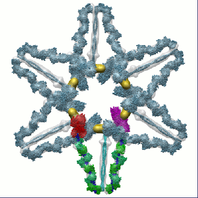
Movie
  • Surface view with section colored by density value
  • Surface level: 0.727346413
  • Imaged by UCSF Chimera
  • Download
  • Surface view colored by cylindrical radius
  • Surface level: 0.727346413
  • Imaged by UCSF Chimera
  • Download
  • Surface view with fitted model
  • Atomic models: PDB-2dfs
  • Surface level: 0.727346413
  • Imaged by UCSF Chimera
  • Download
  • Close-up
  • Surface view with fitted model
  • Atomic models: PDB-2dfs
  • Surface level: 0.727346413
  • Imaged by UCSF Chimera
  • Download
  • Simplified surface model + fitted atomic model
  • Atomic modelsPDB-2dfs
  • Imaged by Jmol
  • Download
Movie viewer
Structure viewerEM map:
SurfViewMolmilJmol/JSmol
Supplemental images

Downloads & links

-
Map

FileDownload / File: emd_1201.map.gz / Format: CCP4 / Size: 7.2 MB / Type: IMAGE STORED AS FLOATING POINT NUMBER (4 BYTES)
AnnotationThis the 3-D average map of myosin-V in "off" state
Voxel sizeX=Y=Z: 5.56 Å
Density
Contour LevelBy EMDB: 0.65 / Movie #1: 0.7273464
Minimum - Maximum-0.411051 - 13.8574
Average (Standard dev.)0.0420816 (±0.651013)
SymmetrySpace group: 1
Details

EMDB XML:

Map geometry
Axis orderXYZ
Origin-90-90-30
Dimensions18018060
Spacing18018060
CellA: 1000.8 Å / B: 1000.8 Å / C: 333.6 Å
α=β=γ: 90 °

CCP4 map header:

modeImage stored as Reals
Å/pix. X/Y/Z5.565.565.56
M x/y/z18018060
origin x/y/z0.0000.0000.000
length x/y/z1000.8001000.800333.600
α/β/γ90.00090.00090.000
start NX/NY/NZ-80-80-79
NX/NY/NZ160160160
MAP C/R/S123
start NC/NR/NS-90-90-30
NC/NR/NS18018060
D min/max/mean-0.41113.8570.042

-
Supplemental data

-
Sample components

-
Entire : MYOSIN V

EntireName: MYOSIN V
Components
  • Sample: MYOSIN V
  • Protein or peptide: Myosin-V

-
Supramolecule #1000: MYOSIN V

SupramoleculeName: MYOSIN V / type: sample / ID: 1000 / Number unique components: 1
Molecular weightExperimental: 400 KDa / Theoretical: 400 KDa

-
Macromolecule #1: Myosin-V

MacromoleculeName: Myosin-V / type: protein_or_peptide / ID: 1 / Name.synonym: MYOV / Details: expressed full lengh myosin-V / Number of copies: 2 / Oligomeric state: Dimer / Recombinant expression: Yes
Source (natural)Organism: Mus musculus (house mouse) / synonym: mouse
Molecular weightExperimental: 400 KDa / Theoretical: 400 KDa
Recombinant expressionOrganism: Sf9 cells

-
Experimental details

-
Structure determination

Methodnegative staining, cryo EM
Processingsubtomogram averaging
Aggregation state2D array

-
Sample preparation

BufferDetails: 20 mM Na2HPO4, 80-100 mM NaCl, 2 mM MgCl2, 1 mM ADP, 1 mM EGTA
StainingType: NEGATIVE
Details: MyoV 2-D arrays were recovered from the lipid monolayer
GridDetails: 200 mesh copper grids covered with a reticulated carbon film.
VitrificationCryogen name: ETHANE / Chamber humidity: 90 % / Chamber temperature: 277 K / Instrument: HOMEMADE PLUNGER / Details: Vitrification instrument: in house plunger / Method: Blot for 3 seconds before plunging
Detailscrystals grown on a lipid-monolayer

-
Electron microscopy

MicroscopeFEI/PHILIPS CM300FEG/ST
Electron beamAcceleration voltage: 300 kV / Electron source: FIELD EMISSION GUN
Electron opticsIllumination mode: FLOOD BEAM / Imaging mode: BRIGHT FIELDBright-field microscopy / Cs: 2.0 mm / Nominal defocus max: 12.0 µm / Nominal defocus min: 4.0 µm / Nominal magnification: 24000
Sample stageSpecimen holder: Eucentric / Specimen holder model: GATAN LIQUID NITROGEN / Tilt series - Axis1 - Min angle: 0 ° / Tilt series - Axis1 - Max angle: 70 °
TemperatureAverage: 103 K
DateFeb 1, 2004
Image recordingCategory: CCD / Film or detector model: TVIPS TEMCAM-F224 (2k x 2k) / Digitization - Sampling interval: 24 µm / Number real images: 360 / Average electron dose: 30 e/Å2

-
Image processing

Crystal parametersUnit cell - A: 653 Å / Unit cell - B: 653 Å / Unit cell - γ: 120 ° / Unit cell - α: 90.0 ° / Unit cell - β: 90.0 ° / Plane group: P 6
CTF correctionDetails: focus gradient correction
Final reconstructionAlgorithm: OTHER / Resolution.type: BY AUTHOR / Resolution: 24.0 Å / Resolution method: FSC 0.5 CUT-OFF / Software - Name: protomo

-
Atomic model buiding 1

SoftwareName: Situs and NMFF
DetailsProtocol: Rigid body and Normal Mode Flexible. Start with Rigid body docking by using SITUS and refine with NMFF
RefinementSpace: REAL / Protocol: FLEXIBLE FIT / Target criteria: Correlation
Output model

PDB-2dfs:
3-D structure of Myosin-V inhibited state

+
About Yorodumi

-
News

-
Feb 9, 2022. New format data for meta-information of EMDB entries

New format data for meta-information of EMDB entries

  • Version 3 of the EMDB header file is now the official format.
  • The previous official version 1.9 will be removed from the archive.

Related info.:EMDB header

External links:wwPDB to switch to version 3 of the EMDB data model

-
Aug 12, 2020. Covid-19 info

Covid-19 info

URL: https://pdbj.org/emnavi/covid19.php

New page: Covid-19 featured information page in EM Navigator.

Related info.:Covid-19 info / Mar 5, 2020. Novel coronavirus structure data

+
Mar 5, 2020. Novel coronavirus structure data

Novel coronavirus structure data

Related info.:Yorodumi Speices / Aug 12, 2020. Covid-19 info

External links:COVID-19 featured content - PDBj / Molecule of the Month (242):Coronavirus Proteases

+
Jan 31, 2019. EMDB accession codes are about to change! (news from PDBe EMDB page)

EMDB accession codes are about to change! (news from PDBe EMDB page)

  • The allocation of 4 digits for EMDB accession codes will soon come to an end. Whilst these codes will remain in use, new EMDB accession codes will include an additional digit and will expand incrementally as the available range of codes is exhausted. The current 4-digit format prefixed with “EMD-” (i.e. EMD-XXXX) will advance to a 5-digit format (i.e. EMD-XXXXX), and so on. It is currently estimated that the 4-digit codes will be depleted around Spring 2019, at which point the 5-digit format will come into force.
  • The EM Navigator/Yorodumi systems omit the EMD- prefix.

Related info.:Q: What is EMD? / ID/Accession-code notation in Yorodumi/EM Navigator

External links:EMDB Accession Codes are Changing Soon! / Contact to PDBj

+
Jul 12, 2017. Major update of PDB

Major update of PDB

  • wwPDB released updated PDB data conforming to the new PDBx/mmCIF dictionary.
  • This is a major update changing the version number from 4 to 5, and with Remediation, in which all the entries are updated.
  • In this update, many items about electron microscopy experimental information are reorganized (e.g. em_software).
  • Now, EM Navigator and Yorodumi are based on the updated data.

External links:wwPDB Remediation / Enriched Model Files Conforming to OneDep Data Standards Now Available in the PDB FTP Archive

-
Yorodumi

Thousand views of thousand structures

  • Yorodumi is a browser for structure data from EMDB, PDB, SASBDB, etc.
  • This page is also the successor to EM Navigator detail page, and also detail information page/front-end page for Omokage search.
  • The word "yorodu" (or yorozu) is an old Japanese word meaning "ten thousand". "mi" (miru) is to see.

Related info.:EMDB / PDB / SASBDB / Comparison of 3 databanks / Yorodumi Search / Aug 31, 2016. New EM Navigator & Yorodumi / Yorodumi Papers / Jmol/JSmol / Function and homology information / Changes in new EM Navigator and Yorodumi

Read more