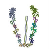
ジャーナル: Nature / 年: 2006 タイトル: Three-dimensional structure of the myosin V inhibited state by cryoelectron tomography. 著者: Jun Liu / Dianne W Taylor / Elena B Krementsova / Kathleen M Trybus / Kenneth A Taylor / 要旨: Unconventional myosin V (myoV) is an actin-based molecular motor that has a key function in organelle and mRNA transport, as well as in membrane trafficking. MyoV was the first member of the myosin ...Unconventional myosin V (myoV) is an actin-based molecular motor that has a key function in organelle and mRNA transport, as well as in membrane trafficking. MyoV was the first member of the myosin superfamily shown to be processive, meaning that a single motor protein can 'walk' hand-over-hand along an actin filament for many steps before detaching. Full-length myoV has a low actin-activated MgATPase activity at low [Ca2+], whereas expressed constructs lacking the cargo-binding domain have a high activity regardless of [Ca2+] (refs 5-7). Hydrodynamic data and electron micrographs indicate that the active state is extended, whereas the inactive state is compact. Here we show the first three-dimensional structure of the myoV inactive state. Each myoV molecule consists of two heads that contain an amino-terminal motor domain followed by a lever arm that binds six calmodulins. The heads are followed by a coiled-coil dimerization domain (S2) and a carboxy-terminal globular cargo-binding domain. In the inactive structure, bending of myoV at the head-S2 junction places the cargo-binding domain near the motor domain's ATP-binding pocket, indicating that ATPase inhibition might occur through decreased rates of nucleotide exchange. The actin-binding interfaces are unobstructed, and the lever arm is oriented in a position typical of strong actin-binding states. This structure indicates that motor recycling after cargo delivery might occur through transport on actively treadmilling actin filaments rather than by diffusion.
詳細: 20 mM Na2HPO4, 80-100 mM NaCl, 2 mM MgCl2, 1 mM ADP, 1 mM EGTA
染色
タイプ: NEGATIVE 詳細: MyoV 2-D arrays were recovered from the lipid monolayer
グリッド
詳細: 200 mesh copper grids covered with a reticulated carbon film.
凍結
凍結剤: ETHANE / チャンバー内湿度: 90 % / チャンバー内温度: 277 K / 装置: HOMEMADE PLUNGER / 詳細: Vitrification instrument: in house plunger / 手法: Blot for 3 seconds before plunging
 ムービー
ムービー コントローラー


 データを開く
 基本情報
基本情報 マップデータ
 試料
試料 機能・相同性情報
 データ登録者
 引用
引用
 構造の表示
構造の表示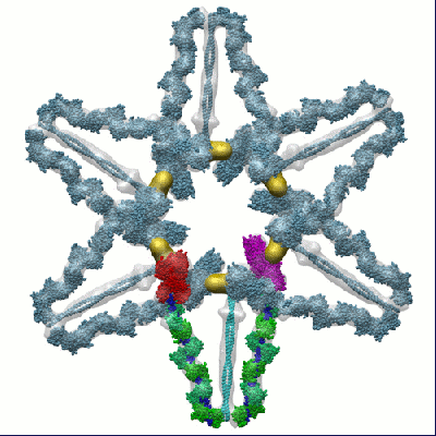
 ダウンロードとリンク
 1201.gif
 http://ftp.pdbj.org/pub/emdb/structures/EMD-1201


 Z (Sec.)
 Y (Row.)
 X (Col.)


 試料の構成要素
 解析
解析 電子顕微鏡法
 FIELD EMISSION GUN