European Union, United Kingdom, Japan, Germany, 5 items
Organization
Grant number
Country
European Research Council (ERC)
ERC-CoG-648432
European Union
Medical Research Council (MRC, United Kingdom)
MC_UP_A025_1013
United Kingdom
Medical Research Council (MRC, United Kingdom)
MC_UP_1201/16
United Kingdom
Japan Society for the Promotion of Science (JSPS)
Japan
German Research Foundation (DFG)
240245660-SFB 1129
Germany
Citation
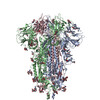
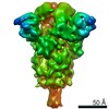
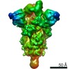
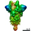
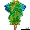
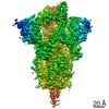
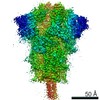
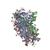
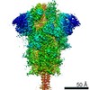
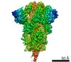
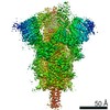
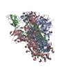
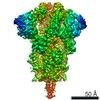
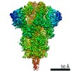
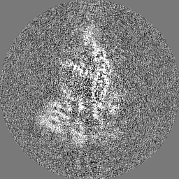
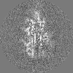
Journal: Nature / Year: 2020 Title: Structures and distributions of SARS-CoV-2 spike proteins on intact virions. Authors: Zunlong Ke / Joaquin Oton / Kun Qu / Mirko Cortese / Vojtech Zila / Lesley McKeane / Takanori Nakane / Jasenko Zivanov / Christopher J Neufeldt / Berati Cerikan / John M Lu / Julia Peukes / ...Authors: Zunlong Ke / Joaquin Oton / Kun Qu / Mirko Cortese / Vojtech Zila / Lesley McKeane / Takanori Nakane / Jasenko Zivanov / Christopher J Neufeldt / Berati Cerikan / John M Lu / Julia Peukes / Xiaoli Xiong / Hans-Georg Kräusslich / Sjors H W Scheres / Ralf Bartenschlager / John A G Briggs / Abstract: Severe acute respiratory syndrome coronavirus 2 (SARS-CoV-2) virions are surrounded by a lipid bilayer from which spike (S) protein trimers protrude. Heavily glycosylated S trimers bind to the ...Severe acute respiratory syndrome coronavirus 2 (SARS-CoV-2) virions are surrounded by a lipid bilayer from which spike (S) protein trimers protrude. Heavily glycosylated S trimers bind to the angiotensin-converting enzyme 2 receptor and mediate entry of virions into target cells. S exhibits extensive conformational flexibility: it modulates exposure of its receptor-binding site and subsequently undergoes complete structural rearrangement to drive fusion of viral and cellular membranes. The structures and conformations of soluble, overexpressed, purified S proteins have been studied in detail using cryo-electron microscopy, but the structure and distribution of S on the virion surface remain unknown. Here we applied cryo-electron microscopy and tomography to image intact SARS-CoV-2 virions and determine the high-resolution structure, conformational flexibility and distribution of S trimers in situ on the virion surface. These results reveal the conformations of S on the virion, and provide a basis from which to understand interactions between S and neutralizing antibodies during infection or vaccination.
History
Deposition
Jul 28, 2020
-
Header (metadata) release
Aug 5, 2020
-
Map release
Aug 5, 2020
-
Update
Dec 30, 2020
-
Current status
Dec 30, 2020
Processing site: PDBe / Status: Released
-
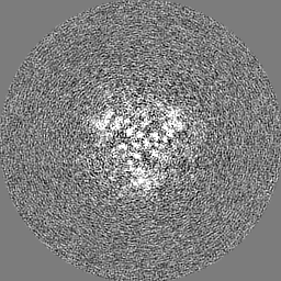
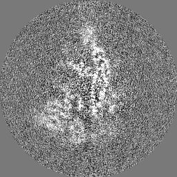
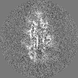
Structure visualization
Movie
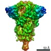
Surface view with section colored by density value
EMPIAR-10492 (Title: Cryo-EM structure of SARS-CoV-2 Spike Proteins on intact virions Data size: 2.1 TB / Data #1: Unaligned movies [micrographs - multiframe])
In the structure databanks used in Yorodumi, some data are registered as the other names, "COVID-19 virus" and "2019-nCoV". Here are the details of the virus and the list of structure data.
Jan 31, 2019. EMDB accession codes are about to change! (news from PDBe EMDB page)
EMDB accession codes are about to change! (news from PDBe EMDB page)
The allocation of 4 digits for EMDB accession codes will soon come to an end. Whilst these codes will remain in use, new EMDB accession codes will include an additional digit and will expand incrementally as the available range of codes is exhausted. The current 4-digit format prefixed with “EMD-” (i.e. EMD-XXXX) will advance to a 5-digit format (i.e. EMD-XXXXX), and so on. It is currently estimated that the 4-digit codes will be depleted around Spring 2019, at which point the 5-digit format will come into force.
The EM Navigator/Yorodumi systems omit the EMD- prefix.
Related info.:Q: What is EMD? / ID/Accession-code notation in Yorodumi/EM Navigator
Yorodumi is a browser for structure data from EMDB, PDB, SASBDB, etc.
This page is also the successor to EM Navigator detail page, and also detail information page/front-end page for Omokage search.
The word "yorodu" (or yorozu) is an old Japanese word meaning "ten thousand". "mi" (miru) is to see.
Related info.:EMDB / PDB / SASBDB / Comparison of 3 databanks / Yorodumi Search / Aug 31, 2016. New EM Navigator & Yorodumi / Yorodumi Papers / Jmol/JSmol / Function and homology information / Changes in new EM Navigator and Yorodumi
 Movie
Movie Controller

 Yorodumi
 Open data
 Basic information
 Map data
 Sample
 Function and homology information
 Severe acute respiratory syndrome coronavirus 2
 Authors
 United Kingdom,
  Japan,
  Germany, 5 items
  Citation
 Structure visualization
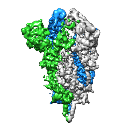
 Downloads & links
 emd_11498.png
 http://ftp.pdbj.org/pub/emdb/structures/EMD-11498
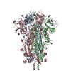


 Z (Sec.)
 Y (Row.)
 X (Col.)


 Sample components
 Processing
 Electron microscopy
 FIELD EMISSION GUN