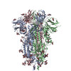
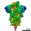
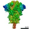
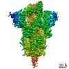
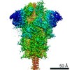
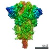
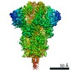
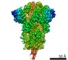
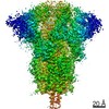
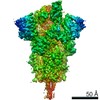
Journal: SSRN / Year: 2020 Title: Structure-Based Design with Tag-Based Purification and In-Process Biotinylation Enable Streamlined Development of SARS-CoV-2 Spike Molecular Probes. Authors: Tongqing Zhou / I-Ting Teng / Adam S Olia / Gabriele Cerutti / Jason Gorman / Alexandra Nazzari / Wei Shi / Yaroslav Tsybovsky / Lingshu Wang / Shuishu Wang / Baoshan Zhang / Yi Zhang / ...Authors: Tongqing Zhou / I-Ting Teng / Adam S Olia / Gabriele Cerutti / Jason Gorman / Alexandra Nazzari / Wei Shi / Yaroslav Tsybovsky / Lingshu Wang / Shuishu Wang / Baoshan Zhang / Yi Zhang / Phinikoula S Katsamba / Yuliya Petrova / Bailey B Banach / Ahmed S Fahad / Lihong Liu / Sheila N Lopez Acevedo / Bharat Madan / Matheus Olivera de Souza / Xiaoli Pan / Pengfei Wang / Jacy R Wolfe / Michael Yin / David D Ho / Emily Phung / Anthony DiPiazza / Lauren Chang / Olubukula Abiona / Kizzmekia S Corbett / Brandon J DeKosky / Barney S Graham / John R Mascola / John Misasi / Tracy Ruckwardt / Nancy J Sullivan / Lawrence Shapiro / Peter D Kwong / Abstract: Biotin-labeled molecular probes, comprising specific regions of the SARS-CoV-2 spike, would be helpful in the isolation and characterization of antibodies targeting this recently emerged pathogen. To ...Biotin-labeled molecular probes, comprising specific regions of the SARS-CoV-2 spike, would be helpful in the isolation and characterization of antibodies targeting this recently emerged pathogen. To develop such probes, we designed constructs incorporating an N-terminal purification tag, a site-specific protease-cleavage site, the probe region of interest, and a C-terminal sequence targeted by biotin ligase. Probe regions included full-length spike ectodomain as well as various subregions, and we also designed mutants to eliminate recognition of the ACE2 receptor. Yields of biotin-labeled probes from transient transfection ranged from ~0.5 mg/L for the complete ectodomain to >5 mg/L for several subregions. Probes were characterized for antigenicity and ACE2 recognition, and the structure of the spike ectodomain probe was determined by cryo-electron microscopy. We also characterized antibody-binding specificities and cell-sorting capabilities of the biotinylated probes. Altogether, structure-based design coupled to efficient purification and biotinylation processes can thus enable streamlined development of SARS-CoV-2 spike-ectodomain probes. Funding: Support for this work was provided by the Intramural Research Program of the Vaccine Research Center, National Institute of Allergy and Infectious Diseases (NIAID). Support for this work was also provided by COVID-19 Fast Grants, the Jack Ma Foundation, the Self Graduate Fellowship Program, and NIH grants DP5OD023118, R21AI143407, and R21AI144408. Some of this work was performed at the Columbia University Cryo-EM Center at the Zuckerman Institute, and some at the Simons Electron Microscopy Center (SEMC) and National Center for Cryo-EM Access and Training (NCCAT) located at the New York Structural Biology Center, supported by grants from the Simons Foundation (SF349247), NYSTAR, and the NIH National Institute of General Medical Sciences (GM103310). Conflict of Interest: The authors declare that they have no conflict of interest. Ethical Approval: Peripheral blood mononuclear cells (PBMCs) for B cell sorting were obtained from a convalescent SARS-CoV-2 patient (collected 75 days post symptom onset under an IRB approved clinical trial protocol, VRC 200 - ClinicalTrials.gov Identifier: NCT00067054) and a healthy control donor from the NIH blood bank pre-SARS-CoV-2 pandemic.
History
Deposition
Jun 15, 2020
-
Header (metadata) release
Sep 2, 2020
-
Map release
Sep 2, 2020
-
Update
May 21, 2025
-
Current status
May 21, 2025
Processing site: RCSB / Status: Released
-
Structure visualization
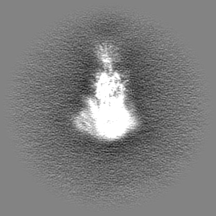
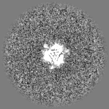
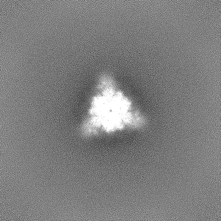
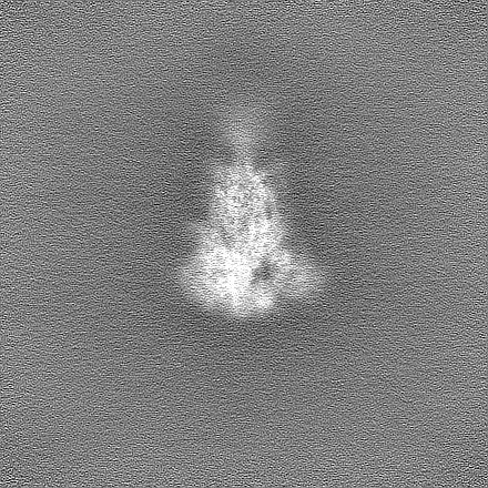
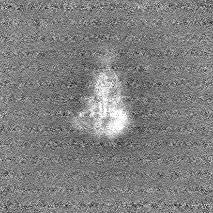
Movie
Surface view with section colored by density value
In the structure databanks used in Yorodumi, some data are registered as the other names, "COVID-19 virus" and "2019-nCoV". Here are the details of the virus and the list of structure data.
Jan 31, 2019. EMDB accession codes are about to change! (news from PDBe EMDB page)
EMDB accession codes are about to change! (news from PDBe EMDB page)
The allocation of 4 digits for EMDB accession codes will soon come to an end. Whilst these codes will remain in use, new EMDB accession codes will include an additional digit and will expand incrementally as the available range of codes is exhausted. The current 4-digit format prefixed with “EMD-” (i.e. EMD-XXXX) will advance to a 5-digit format (i.e. EMD-XXXXX), and so on. It is currently estimated that the 4-digit codes will be depleted around Spring 2019, at which point the 5-digit format will come into force.
The EM Navigator/Yorodumi systems omit the EMD- prefix.
Related info.:Q: What is EMD? / ID/Accession-code notation in Yorodumi/EM Navigator
Yorodumi is a browser for structure data from EMDB, PDB, SASBDB, etc.
This page is also the successor to EM Navigator detail page, and also detail information page/front-end page for Omokage search.
The word "yorodu" (or yorozu) is an old Japanese word meaning "ten thousand". "mi" (miru) is to see.
Related info.:EMDB / PDB / SASBDB / Comparison of 3 databanks / Yorodumi Search / Aug 31, 2016. New EM Navigator & Yorodumi / Yorodumi Papers / Jmol/JSmol / Function and homology information / Changes in new EM Navigator and Yorodumi
 Movie
Movie Controller

 Yorodumi
 Open data
 Basic information
 Map data
 Sample
 Keywords
 Function and homology information
 Severe acute respiratory syndrome coronavirus 2
 Authors
 Citation
 Structure visualization
 Downloads & links
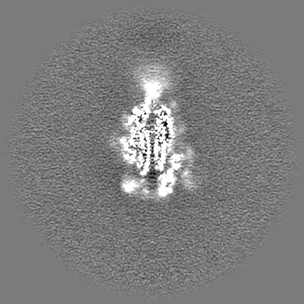
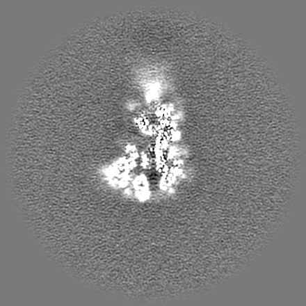
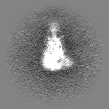
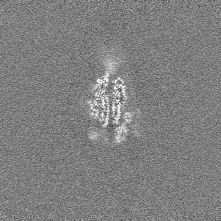
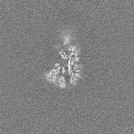
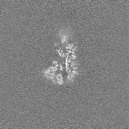
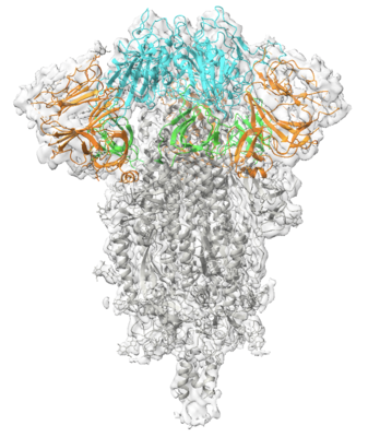 emd_22161.png
 http://ftp.pdbj.org/pub/emdb/structures/EMD-22161


 Z (Sec.)
 Y (Row.)
 X (Col.)


 Sample components
 Homo sapiens (human)
 Processing
 Electron microscopy
 FIELD EMISSION GUN