[English] 日本語
Yorodumi
- EMDB-11207: Furin Cleaved Spike Protein of SARS-CoV-2 in Closed Conformation -

+
Open data


ID or keywords:

Loading...

-
Basic information

Entry
Database: EMDB / ID: EMD-11207
TitleFurin Cleaved Spike Protein of SARS-CoV-2 in Closed Conformation
Map data
Sample
  • Complex: SARS-CoV-2 Spike Glycoprotein Ectodomain
    • Protein or peptide: Spike glycoproteinSpike protein
  • Ligand: 2-acetamido-2-deoxy-beta-D-glucopyranose
Function / homology
Function and homology information


Maturation of spike protein / viral translation / Translation of Structural Proteins / Virion Assembly and Release / host cell surface / host extracellular space / suppression by virus of host tetherin activity / Induction of Cell-Cell Fusion / structural constituent of virion / host cell endoplasmic reticulum-Golgi intermediate compartment membrane ...Maturation of spike protein / viral translation / Translation of Structural Proteins / Virion Assembly and Release / host cell surface / host extracellular space / suppression by virus of host tetherin activity / Induction of Cell-Cell Fusion / structural constituent of virion / host cell endoplasmic reticulum-Golgi intermediate compartment membrane / entry receptor-mediated virion attachment to host cell / receptor-mediated endocytosis of virus by host cell / Attachment and Entry / membrane fusion / positive regulation of viral entry into host cell / receptor-mediated virion attachment to host cell / receptor ligand activity / host cell surface receptor binding / fusion of virus membrane with host plasma membrane / fusion of virus membrane with host endosome membrane / viral envelope / symbiont-mediated suppression of host type I interferon-mediated signaling pathway / virion attachment to host cell / SARS-CoV-2 activates/modulates innate and adaptive immune responses / host cell plasma membrane / virion membrane / identical protein binding / plasma membrane
Similarity search - Function
Spike (S) protein S1 subunit, receptor-binding domain, SARS-CoV-2 / Spike (S) protein S1 subunit, N-terminal domain, SARS-CoV-like / Betacoronavirus spike (S) glycoprotein S1 subunit N-terminal (NTD) domain profile. / Spike glycoprotein, N-terminal domain superfamily / Betacoronavirus spike (S) glycoprotein S1 subunit C-terminal (CTD) domain profile. / Spike glycoprotein, betacoronavirus / Spike (S) protein S1 subunit, receptor-binding domain, betacoronavirus / Spike S1 subunit, receptor binding domain superfamily, betacoronavirus / Betacoronavirus spike glycoprotein S1, receptor binding / Spike glycoprotein S1, N-terminal domain, betacoronavirus-like ...Spike (S) protein S1 subunit, receptor-binding domain, SARS-CoV-2 / Spike (S) protein S1 subunit, N-terminal domain, SARS-CoV-like / Betacoronavirus spike (S) glycoprotein S1 subunit N-terminal (NTD) domain profile. / Spike glycoprotein, N-terminal domain superfamily / Betacoronavirus spike (S) glycoprotein S1 subunit C-terminal (CTD) domain profile. / Spike glycoprotein, betacoronavirus / Spike (S) protein S1 subunit, receptor-binding domain, betacoronavirus / Spike S1 subunit, receptor binding domain superfamily, betacoronavirus / Betacoronavirus spike glycoprotein S1, receptor binding / Spike glycoprotein S1, N-terminal domain, betacoronavirus-like / Betacoronavirus-like spike glycoprotein S1, N-terminal / Spike glycoprotein S2, coronavirus, heptad repeat 1 / Spike glycoprotein S2, coronavirus, heptad repeat 2 / Coronavirus spike (S) glycoprotein S2 subunit heptad repeat 2 (HR2) region profile. / Coronavirus spike (S) glycoprotein S2 subunit heptad repeat 1 (HR1) region profile. / Spike glycoprotein S2 superfamily, coronavirus / Spike glycoprotein S2, coronavirus / Coronavirus spike glycoprotein S2 / Coronavirus spike glycoprotein S1, C-terminal / Coronavirus spike glycoprotein S1, C-terminal
Similarity search - Domain/homology
Biological speciesSevere acute respiratory syndrome coronavirus 2
Methodsingle particle reconstruction / cryo EM / Resolution: 2.9 Å
AuthorsWrobel AG / Benton DJ / Rosenthal PB / Gamblin SJ
Funding support United Kingdom, 2 items
OrganizationGrant numberCountry
The Francis Crick InstituteFC001078 United Kingdom
The Francis Crick InstituteFC001143 United Kingdom
Citation
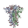
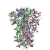
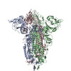
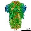
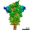
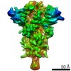
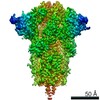
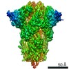
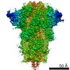
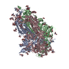
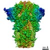
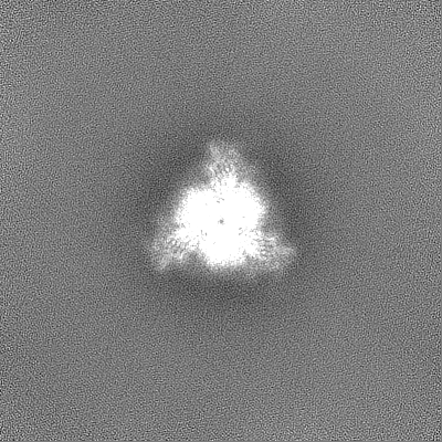
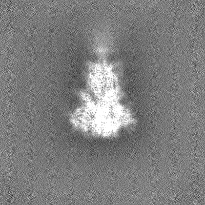
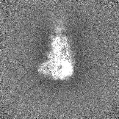
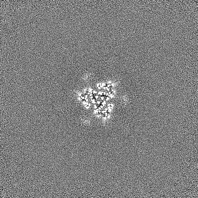
Journal: Nat Struct Mol Biol / Year: 2020
Title: SARS-CoV-2 and bat RaTG13 spike glycoprotein structures inform on virus evolution and furin-cleavage effects.
Authors: Antoni G Wrobel / Donald J Benton / Pengqi Xu / Chloë Roustan / Stephen R Martin / Peter B Rosenthal / John J Skehel / Steven J Gamblin /
Abstract: SARS-CoV-2 is thought to have emerged from bats, possibly via a secondary host. Here, we investigate the relationship of spike (S) glycoprotein from SARS-CoV-2 with the S protein of a closely related ...SARS-CoV-2 is thought to have emerged from bats, possibly via a secondary host. Here, we investigate the relationship of spike (S) glycoprotein from SARS-CoV-2 with the S protein of a closely related bat virus, RaTG13. We determined cryo-EM structures for RaTG13 S and for both furin-cleaved and uncleaved SARS-CoV-2 S; we compared these with recently reported structures for uncleaved SARS-CoV-2 S. We also biochemically characterized their relative stabilities and affinities for the SARS-CoV-2 receptor ACE2. Although the overall structures of human and bat virus S proteins are similar, there are key differences in their properties, including a more stable precleavage form of human S and about 1,000-fold tighter binding of SARS-CoV-2 to human receptor. These observations suggest that cleavage at the furin-cleavage site decreases the overall stability of SARS-CoV-2 S and facilitates the adoption of the open conformation that is required for S to bind to the ACE2 receptor.
History
DepositionJun 18, 2020-
Header (metadata) releaseJul 1, 2020-
Map releaseJul 1, 2020-
UpdateSep 16, 2020-
Current statusSep 16, 2020Processing site: PDBe / Status: Released

-
Structure visualization

Movie
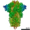
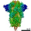
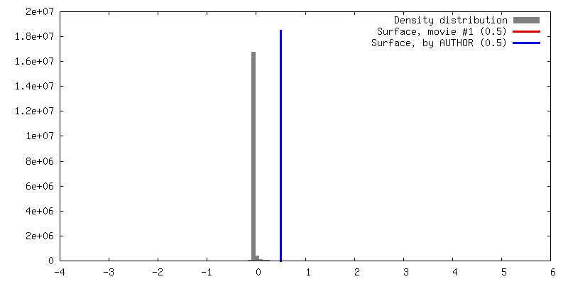
  • Surface view with section colored by density value
  • Surface level: 0.5
  • Imaged by UCSF Chimera
  • Download
  • Surface view colored by cylindrical radius
  • Surface level: 0.5
  • Imaged by UCSF Chimera
  • Download
  • Surface view with fitted model
  • Atomic models: PDB-6zgi
  • Surface level: 0.6
  • Imaged by UCSF Chimera
  • Download
Movie viewer
Structure viewerEM map:
SurfViewMolmilJmol/JSmol
Supplemental images

Downloads & links

-
Map

FileDownload / File: emd_11207.map.gz / Format: CCP4 / Size: 244.1 MB / Type: IMAGE STORED AS FLOATING POINT NUMBER (4 BYTES)
Voxel sizeX=Y=Z: 1.078 Å
Density
Contour LevelBy AUTHOR: 0.5 / Movie #1: 0.5
Minimum - Maximum-3.7558324 - 5.4638
Average (Standard dev.)0.0027707093 (±0.08639582)
SymmetrySpace group: 1
Details

EMDB XML:

Map geometry
Axis orderXYZ
Origin000
Dimensions400400400
Spacing400400400
CellA=B=C: 431.19998 Å
α=β=γ: 90.0 °

CCP4 map header:

modeImage stored as Reals
Å/pix. X/Y/Z1.0781.0781.078
M x/y/z400400400
origin x/y/z0.0000.0000.000
length x/y/z431.200431.200431.200
α/β/γ90.00090.00090.000
MAP C/R/S123
start NC/NR/NS000
NC/NR/NS400400400
D min/max/mean-3.7565.4640.003

-
Supplemental data

-
Mask #1

Fileemd_11207_msk_1.map
Projections & Slices
AxesZYX

Projections

Slices (1/2)
Density Histograms

-
Half map: #2

Fileemd_11207_half_map_1.map
Projections & Slices
AxesZYX

Projections

Slices (1/2)
Density Histograms

-
Half map: #1

Fileemd_11207_half_map_2.map
Projections & Slices
AxesZYX

Projections

Slices (1/2)
Density Histograms

-
Sample components

-
Entire : SARS-CoV-2 Spike Glycoprotein Ectodomain

EntireName: SARS-CoV-2 Spike Glycoprotein Ectodomain
Components
  • Complex: SARS-CoV-2 Spike Glycoprotein Ectodomain
    • Protein or peptide: Spike glycoproteinSpike protein
  • Ligand: 2-acetamido-2-deoxy-beta-D-glucopyranose

-
Supramolecule #1: SARS-CoV-2 Spike Glycoprotein Ectodomain

SupramoleculeName: SARS-CoV-2 Spike Glycoprotein Ectodomain / type: complex / ID: 1 / Parent: 0 / Macromolecule list: #1
Source (natural)Organism: Severe acute respiratory syndrome coronavirus 2
Recombinant expressionOrganism: Homo sapiens (human)
Molecular weightTheoretical: 500 KDa

-
Macromolecule #1: Spike glycoprotein

MacromoleculeName: Spike glycoprotein / type: protein_or_peptide / ID: 1 / Number of copies: 3 / Enantiomer: LEVO
Source (natural)Organism: Severe acute respiratory syndrome coronavirus 2
Molecular weightTheoretical: 142.410078 KDa
Recombinant expressionOrganism: Homo sapiens (human)
SequenceString: MGILPSPGMP ALLSLVSLLS VLLMGCVAET GMFVFLVLLP LVSSQCVNLT TRTQLPPAYT NSFTRGVYYP DKVFRSSVLH STQDLFLPF FSNVTWFHAI HVSGTNGTKR FDNPVLPFND GVYFASTEKS NIIRGWIFGT TLDSKTQSLL IVNNATNVVI K VCEFQFCN ...String:
MGILPSPGMP ALLSLVSLLS VLLMGCVAET GMFVFLVLLP LVSSQCVNLT TRTQLPPAYT NSFTRGVYYP DKVFRSSVLH STQDLFLPF FSNVTWFHAI HVSGTNGTKR FDNPVLPFND GVYFASTEKS NIIRGWIFGT TLDSKTQSLL IVNNATNVVI K VCEFQFCN DPFLGVYYHK NNKSWMESEF RVYSSANNCT FEYVSQPFLM DLEGKQGNFK NLREFVFKNI DGYFKIYSKH TP INLVRDL PQGFSALEPL VDLPIGINIT RFQTLLALHR SYLTPGDSSS GWTAGAAAYY VGYLQPRTFL LKYNENGTIT DAV DCALDP LSETKCTLKS FTVEKGIYQT SNFRVQPTES IVRFPNITNL CPFGEVFNAT RFASVYAWNR KRISNCVADY SVLY NSASF STFKCYGVSP TKLNDLCFTN VYADSFVIRG DEVRQIAPGQ TGKIADYNYK LPDDFTGCVI AWNSNNLDSK VGGNY NYLY RLFRKSNLKP FERDISTEIY QAGSTPCNGV EGFNCYFPLQ SYGFQPTNGV GYQPYRVVVL SFELLHAPAT VCGPKK STN LVKNKCVNFN FNGLTGTGVL TESNKKFLPF QQFGRDIADT TDAVRDPQTL EILDITPCSF GGVSVITPGT NTSNQVA VL YQDVNCTEVP VAIHADQLTP TWRVYSTGSN VFQTRAGCLI GAEHVNNSYE CDIPIGAGIC ASYQTQTNSP RRARSVAS Q SIIAYTMSLG AENSVAYSNN SIAIPTNFTI SVTTEILPVS MTKTSVDCTM YICGDSTECS NLLLQYGSFC TQLNRALTG IAVEQDKNTQ EVFAQVKQIY KTPPIKDFGG FNFSQILPDP SKPSKRSFIE DLLFNKVTLA DAGFIKQYGD CLGDIAARDL ICAQKFNGL TVLPPLLTDE MIAQYTSALL AGTITSGWTF GAGAALQIPF AMQMAYRFNG IGVTQNVLYE NQKLIANQFN S AIGKIQDS LSSTASALGK LQDVVNQNAQ ALNTLVKQLS SNFGAISSVL NDILSRLDPP EAEVQIDRLI TGRLQSLQTY VT QQLIRAA EIRASANLAA TKMSECVLGQ SKRVDFCGKG YHLMSFPQSA PHGVVFLHVT YVPAQEKNFT TAPAICHDGK AHF PREGVF VSNGTHWFVT QRNFYEPQII TTDNTFVSGN CDVVIGIVNN TVYDPLQPEL DSFKEELDKY FKNHTSPDVD LGDI SGINA SVVNIQKEID RLNEVAKNLN ESLIDLQELG KYEQSGRENL YFQGGGGSGY IPEAPRDGQA YVRKDGEWVL LSTFL GHHH HHH

-
Macromolecule #3: 2-acetamido-2-deoxy-beta-D-glucopyranose

MacromoleculeName: 2-acetamido-2-deoxy-beta-D-glucopyranose / type: ligand / ID: 3 / Number of copies: 24 / Formula: NAG
Molecular weightTheoretical: 221.208 Da
Chemical component information

ChemComp-NAG:
2-acetamido-2-deoxy-beta-D-glucopyranose / N-Acetylglucosamine

-
Experimental details

-
Structure determination

Methodcryo EM
Processingsingle particle reconstruction
Aggregation stateparticle

-
Sample preparation

Concentration0.1 mg/mL
BufferpH: 8
GridModel: Quantifoil R2/2 / Material: COPPER / Mesh: 200 / Support film - Material: CARBON / Support film - topology: CONTINUOUS
VitrificationCryogen name: ETHANE / Chamber humidity: 100 % / Chamber temperature: 277 K / Instrument: FEI VITROBOT MARK III

-
Electron microscopy

MicroscopeFEI TITAN KRIOS
Electron beamAcceleration voltage: 300 kV / Electron source: FIELD EMISSION GUN
Electron opticsIllumination mode: FLOOD BEAM / Imaging mode: BRIGHT FIELDBright-field microscopy / Nominal defocus max: 3.0 µm / Nominal defocus min: 1.5 µm
Specialist opticsEnergy filter - Name: GIF Quantum LS / Energy filter - Slit width: 20 eV
Sample stageSpecimen holder model: FEI TITAN KRIOS AUTOGRID HOLDER / Cooling holder cryogen: NITROGEN
Image recordingFilm or detector model: GATAN K2 QUANTUM (4k x 4k) / Detector mode: COUNTING / Average exposure time: 8.0 sec. / Average electron dose: 54.4 e/Å2
Experimental equipment
Model: Titan Krios / Image courtesy: FEI Company

-
Image processing

CTF correctionSoftware - Name: CTFFIND (ver. 4.1.10)
Startup modelType of model: INSILICO MODEL / In silico model: cryoSPARC Ab Initio
Initial angle assignmentType: MAXIMUM LIKELIHOOD
Final angle assignmentType: MAXIMUM LIKELIHOOD / Software - Name: cryoSPARC (ver. 2.15)
Final reconstructionApplied symmetry - Point group: C3 (3 fold cyclic) / Algorithm: FOURIER SPACE / Resolution.type: BY AUTHOR / Resolution: 2.9 Å / Resolution method: FSC 0.143 CUT-OFF / Software - Name: cryoSPARC (ver. 2.15) / Number images used: 95000
FSC plot (resolution estimation)

-
Atomic model buiding 1

Initial modelPDB ID:

Chain - Chain ID: A
RefinementSpace: REAL / Protocol: OTHER
Output model

PDB-6zgi:
Furin Cleaved Spike Protein of SARS-CoV-2 in Closed Conformation

+
About Yorodumi

-
News

-
Feb 9, 2022. New format data for meta-information of EMDB entries

New format data for meta-information of EMDB entries

  • Version 3 of the EMDB header file is now the official format.
  • The previous official version 1.9 will be removed from the archive.

Related info.:EMDB header

External links:wwPDB to switch to version 3 of the EMDB data model

-
Aug 12, 2020. Covid-19 info

Covid-19 info

URL: https://pdbj.org/emnavi/covid19.php

New page: Covid-19 featured information page in EM Navigator.

Related info.:Covid-19 info / Mar 5, 2020. Novel coronavirus structure data

+
Mar 5, 2020. Novel coronavirus structure data

Novel coronavirus structure data

Related info.:Yorodumi Speices / Aug 12, 2020. Covid-19 info

External links:COVID-19 featured content - PDBj / Molecule of the Month (242):Coronavirus Proteases

+
Jan 31, 2019. EMDB accession codes are about to change! (news from PDBe EMDB page)

EMDB accession codes are about to change! (news from PDBe EMDB page)

  • The allocation of 4 digits for EMDB accession codes will soon come to an end. Whilst these codes will remain in use, new EMDB accession codes will include an additional digit and will expand incrementally as the available range of codes is exhausted. The current 4-digit format prefixed with “EMD-” (i.e. EMD-XXXX) will advance to a 5-digit format (i.e. EMD-XXXXX), and so on. It is currently estimated that the 4-digit codes will be depleted around Spring 2019, at which point the 5-digit format will come into force.
  • The EM Navigator/Yorodumi systems omit the EMD- prefix.

Related info.:Q: What is EMD? / ID/Accession-code notation in Yorodumi/EM Navigator

External links:EMDB Accession Codes are Changing Soon! / Contact to PDBj

+
Jul 12, 2017. Major update of PDB

Major update of PDB

  • wwPDB released updated PDB data conforming to the new PDBx/mmCIF dictionary.
  • This is a major update changing the version number from 4 to 5, and with Remediation, in which all the entries are updated.
  • In this update, many items about electron microscopy experimental information are reorganized (e.g. em_software).
  • Now, EM Navigator and Yorodumi are based on the updated data.

External links:wwPDB Remediation / Enriched Model Files Conforming to OneDep Data Standards Now Available in the PDB FTP Archive

-
Yorodumi

Thousand views of thousand structures

  • Yorodumi is a browser for structure data from EMDB, PDB, SASBDB, etc.
  • This page is also the successor to EM Navigator detail page, and also detail information page/front-end page for Omokage search.
  • The word "yorodu" (or yorozu) is an old Japanese word meaning "ten thousand". "mi" (miru) is to see.

Related info.:EMDB / PDB / SASBDB / Comparison of 3 databanks / Yorodumi Search / Aug 31, 2016. New EM Navigator & Yorodumi / Yorodumi Papers / Jmol/JSmol / Function and homology information / Changes in new EM Navigator and Yorodumi

Read more