National Institutes of Health/National Institute Of Allergy and Infectious Diseases (NIH/NIAID)
UM1 AI144462
United States
Citation
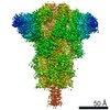
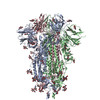
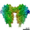
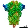
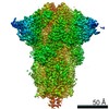
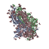
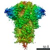
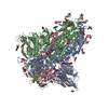
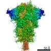
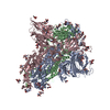
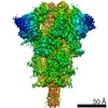
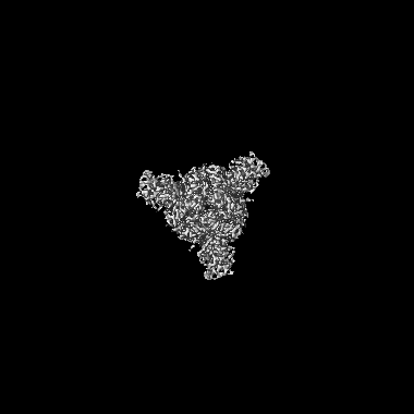
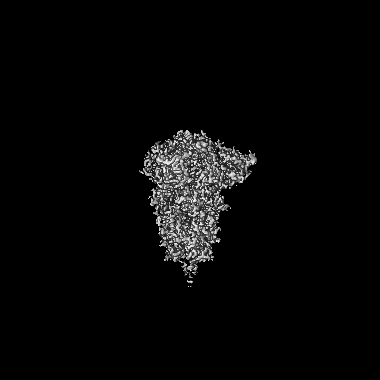
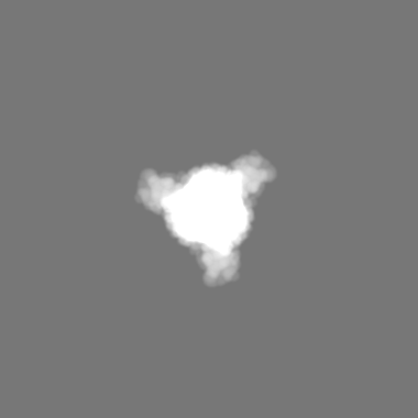
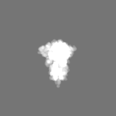
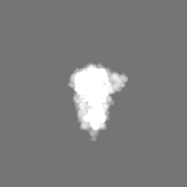
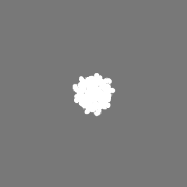
Journal: bioRxiv / Year: 2020 Title: Structural analysis of full-length SARS-CoV-2 spike protein from an advanced vaccine candidate. Authors: Sandhya Bangaru / Gabriel Ozorowski / Hannah L Turner / Aleksandar Antanasijevic / Deli Huang / Xiaoning Wang / Jonathan L Torres / Jolene K Diedrich / Jing-Hui Tian / Alyse D Portnoff / ...Authors: Sandhya Bangaru / Gabriel Ozorowski / Hannah L Turner / Aleksandar Antanasijevic / Deli Huang / Xiaoning Wang / Jonathan L Torres / Jolene K Diedrich / Jing-Hui Tian / Alyse D Portnoff / Nita Patel / Michael J Massare / John R Yates / David Nemazee / James C Paulson / Greg Glenn / Gale Smith / Andrew B Ward / Abstract: Vaccine efforts against the severe acute respiratory syndrome coronavirus 2 (SARS-CoV-2) responsible for the current COVID-19 pandemic are focused on SARS-CoV-2 spike glycoprotein, the primary target ...Vaccine efforts against the severe acute respiratory syndrome coronavirus 2 (SARS-CoV-2) responsible for the current COVID-19 pandemic are focused on SARS-CoV-2 spike glycoprotein, the primary target for neutralizing antibodies. Here, we performed cryo-EM and site-specific glycan analysis of one of the leading subunit vaccine candidates from Novavax based on a full-length spike protein formulated in polysorbate 80 (PS 80) detergent. Our studies reveal a stable prefusion conformation of the spike immunogen with slight differences in the S1 subunit compared to published spike ectodomain structures. Interestingly, we also observed novel interactions between the spike trimers allowing formation of higher order spike complexes. This study confirms the structural integrity of the full-length spike protein immunogen and provides a basis for interpreting immune responses to this multivalent nanoparticle immunogen.
History
Deposition
Jul 26, 2020
-
Header (metadata) release
Aug 26, 2020
-
Map release
Aug 26, 2020
-
Update
Nov 13, 2024
-
Current status
Nov 13, 2024
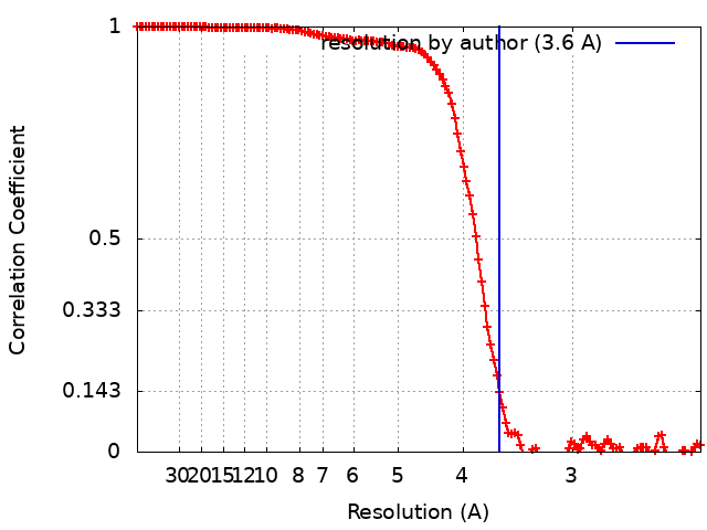
Processing site: RCSB / Status: Released
-
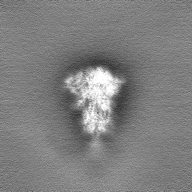
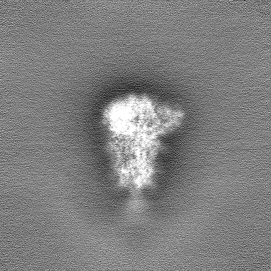
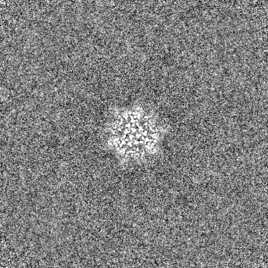
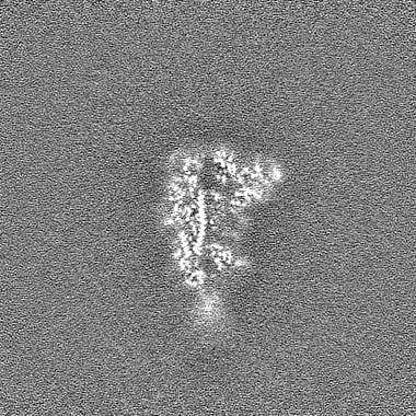
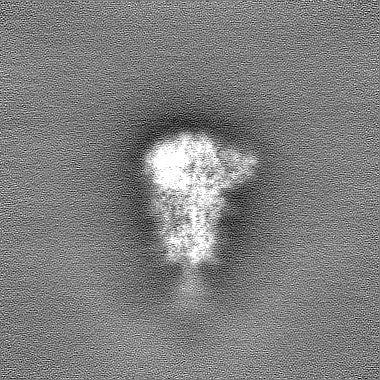
Structure visualization
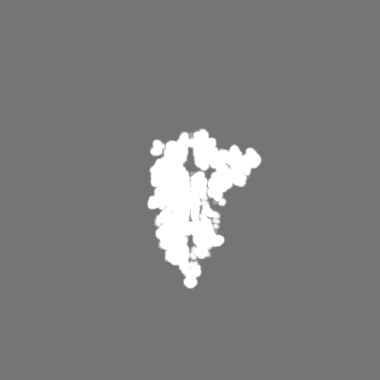
Movie
Surface view with section colored by density value
Model: UltrAuFoil R1.2/1.3 / Material: GOLD / Mesh: 300 / Support film - Material: GOLD / Support film - topology: HOLEY / Pretreatment - Type: PLASMA CLEANING / Pretreatment - Time: 7 sec.
Vitrification
Cryogen name: ETHANE / Chamber humidity: 100 % / Chamber temperature: 283 K / Instrument: FEI VITROBOT MARK IV
-
Electron microscopy
Microscope
FEI TALOS ARCTICA
Image recording
Film or detector model: GATAN K2 SUMMIT (4k x 4k) / Detector mode: COUNTING / Number real images: 5506 / Average exposure time: 11.5 sec. / Average electron dose: 50.0 e/Å2
Electron beam
Acceleration voltage: 200 kV / Electron source: FIELD EMISSION GUN
In the structure databanks used in Yorodumi, some data are registered as the other names, "COVID-19 virus" and "2019-nCoV". Here are the details of the virus and the list of structure data.
Jan 31, 2019. EMDB accession codes are about to change! (news from PDBe EMDB page)
EMDB accession codes are about to change! (news from PDBe EMDB page)
The allocation of 4 digits for EMDB accession codes will soon come to an end. Whilst these codes will remain in use, new EMDB accession codes will include an additional digit and will expand incrementally as the available range of codes is exhausted. The current 4-digit format prefixed with “EMD-” (i.e. EMD-XXXX) will advance to a 5-digit format (i.e. EMD-XXXXX), and so on. It is currently estimated that the 4-digit codes will be depleted around Spring 2019, at which point the 5-digit format will come into force.
The EM Navigator/Yorodumi systems omit the EMD- prefix.
Related info.:Q: What is EMD? / ID/Accession-code notation in Yorodumi/EM Navigator
Yorodumi is a browser for structure data from EMDB, PDB, SASBDB, etc.
This page is also the successor to EM Navigator detail page, and also detail information page/front-end page for Omokage search.
The word "yorodu" (or yorozu) is an old Japanese word meaning "ten thousand". "mi" (miru) is to see.
Related info.:EMDB / PDB / SASBDB / Comparison of 3 databanks / Yorodumi Search / Aug 31, 2016. New EM Navigator & Yorodumi / Yorodumi Papers / Jmol/JSmol / Function and homology information / Changes in new EM Navigator and Yorodumi
 Movie
Movie Controller

 Yorodumi
 Open data
 Basic information
 Map data
 Sample
 Keywords
 Function and homology information
 Severe acute respiratory syndrome coronavirus 2
 Authors
 United States, 2 items
  Citation
 Structure visualization
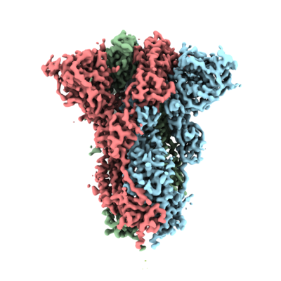
 Downloads & links
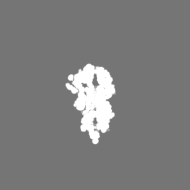
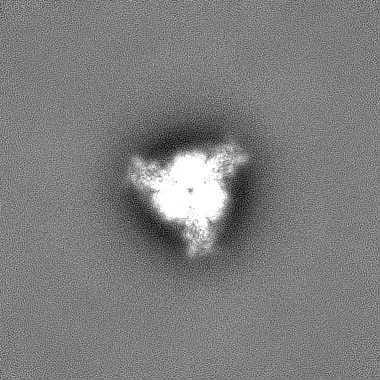
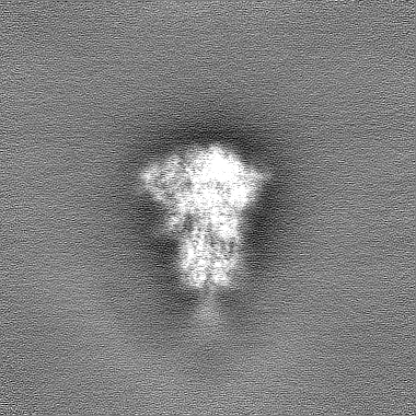
 emd_22352.png
 http://ftp.pdbj.org/pub/emdb/structures/EMD-22352


 Z (Sec.)
 Y (Row.)
 X (Col.)


 Sample components
 Spodoptera frugiperda (fall armyworm)

 Processing
 Electron microscopy
 FIELD EMISSION GUN