Movie
Movie Controller

[English] 日本語
 Yorodumi
- PDB-7jji: Structure of SARS-CoV-2 3Q-2P full-length prefusion spike trimer ... -
+ Open data

- Basic information
| Entry | Database: PDB / ID: 7jji | |||||||||
|---|---|---|---|---|---|---|---|---|---|---|
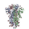
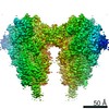
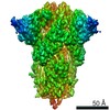
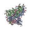
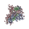
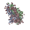
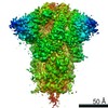
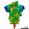
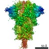
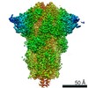
| Title | Structure of SARS-CoV-2 3Q-2P full-length prefusion spike trimer (C3 symmetry) | |||||||||
 Components Components | Spike glycoprotein | |||||||||
 Keywords Keywords | VIRAL PROTEIN / SARS-CoV-2 / Glycoprotein / Immunogen / vaccine | |||||||||
| Function / homology |  Function and homology information Function and homology informationsymbiont-mediated disruption of host tissue / Maturation of spike protein / Translation of Structural Proteins / Virion Assembly and Release / host cell surface / host extracellular region / symbiont-mediated-mediated suppression of host tetherin activity / Induction of Cell-Cell Fusion / structural constituent of virion / positive regulation of viral entry into host cell ...symbiont-mediated disruption of host tissue / Maturation of spike protein / Translation of Structural Proteins / Virion Assembly and Release / host cell surface / host extracellular region / symbiont-mediated-mediated suppression of host tetherin activity / Induction of Cell-Cell Fusion / structural constituent of virion / positive regulation of viral entry into host cell / membrane fusion / host cell endoplasmic reticulum-Golgi intermediate compartment membrane / Attachment and Entry / entry receptor-mediated virion attachment to host cell / receptor-mediated virion attachment to host cell / host cell surface receptor binding / symbiont-mediated suppression of host innate immune response / endocytosis involved in viral entry into host cell / receptor ligand activity / fusion of virus membrane with host plasma membrane / fusion of virus membrane with host endosome membrane / viral envelope / symbiont entry into host cell / virion attachment to host cell / host cell plasma membrane / SARS-CoV-2 activates/modulates innate and adaptive immune responses / virion membrane / membrane / identical protein binding / plasma membrane Similarity search - Function | |||||||||
| Biological species |   Severe acute respiratory syndrome coronavirus 2 Severe acute respiratory syndrome coronavirus 2 | |||||||||
| Method | ELECTRON MICROSCOPY / single particle reconstruction / cryo EM / Resolution: 3.6 Å | |||||||||
 Authors Authors | Bangaru, S. / Turner, H.L. / Ozorowski, G. / Antanasijevic, A. / Ward, A.B. | |||||||||
| Funding support |  United States, 2items United States, 2items
| |||||||||
 Citation Citation | Journal: Science / Year: 2020 Title: Structural analysis of full-length SARS-CoV-2 spike protein from an advanced vaccine candidate. Authors: Sandhya Bangaru / Gabriel Ozorowski / Hannah L Turner / Aleksandar Antanasijevic / Deli Huang / Xiaoning Wang / Jonathan L Torres / Jolene K Diedrich / Jing-Hui Tian / Alyse D Portnoff / ...Authors: Sandhya Bangaru / Gabriel Ozorowski / Hannah L Turner / Aleksandar Antanasijevic / Deli Huang / Xiaoning Wang / Jonathan L Torres / Jolene K Diedrich / Jing-Hui Tian / Alyse D Portnoff / Nita Patel / Michael J Massare / John R Yates / David Nemazee / James C Paulson / Greg Glenn / Gale Smith / Andrew B Ward / Abstract: Vaccine efforts to combat the severe acute respiratory syndrome coronavirus 2 (SARS-CoV-2), which is responsible for the current coronavirus disease 2019 (COVID-19) pandemic, are focused on SARS-CoV- ...Vaccine efforts to combat the severe acute respiratory syndrome coronavirus 2 (SARS-CoV-2), which is responsible for the current coronavirus disease 2019 (COVID-19) pandemic, are focused on SARS-CoV-2 spike glycoprotein, the primary target for neutralizing antibodies. We performed cryo-election microscopy and site-specific glycan analysis of one of the leading subunit vaccine candidates from Novavax, which is based on a full-length spike protein formulated in polysorbate 80 detergent. Our studies reveal a stable prefusion conformation of the spike immunogen with slight differences in the S1 subunit compared with published spike ectodomain structures. We also observed interactions between the spike trimers, allowing formation of higher-order spike complexes. This study confirms the structural integrity of the full-length spike protein immunogen and provides a basis for interpreting immune responses to this multivalent nanoparticle immunogen. #1: Journal: bioRxiv / Year: 2020 Title: Structural analysis of full-length SARS-CoV-2 spike protein from an advanced vaccine candidate. Authors: Sandhya Bangaru / Gabriel Ozorowski / Hannah L Turner / Aleksandar Antanasijevic / Deli Huang / Xiaoning Wang / Jonathan L Torres / Jolene K Diedrich / Jing-Hui Tian / Alyse D Portnoff / ...Authors: Sandhya Bangaru / Gabriel Ozorowski / Hannah L Turner / Aleksandar Antanasijevic / Deli Huang / Xiaoning Wang / Jonathan L Torres / Jolene K Diedrich / Jing-Hui Tian / Alyse D Portnoff / Nita Patel / Michael J Massare / John R Yates / David Nemazee / James C Paulson / Greg Glenn / Gale Smith / Andrew B Ward / Abstract: Vaccine efforts against the severe acute respiratory syndrome coronavirus 2 (SARS-CoV-2) responsible for the current COVID-19 pandemic are focused on SARS-CoV-2 spike glycoprotein, the primary target ...Vaccine efforts against the severe acute respiratory syndrome coronavirus 2 (SARS-CoV-2) responsible for the current COVID-19 pandemic are focused on SARS-CoV-2 spike glycoprotein, the primary target for neutralizing antibodies. Here, we performed cryo-EM and site-specific glycan analysis of one of the leading subunit vaccine candidates from Novavax based on a full-length spike protein formulated in polysorbate 80 (PS 80) detergent. Our studies reveal a stable prefusion conformation of the spike immunogen with slight differences in the S1 subunit compared to published spike ectodomain structures. Interestingly, we also observed novel interactions between the spike trimers allowing formation of higher order spike complexes. This study confirms the structural integrity of the full-length spike protein immunogen and provides a basis for interpreting immune responses to this multivalent nanoparticle immunogen. | |||||||||
| History |
|
- Structure visualization
| Movie |
Movie viewer |
|---|---|
| Structure viewer | Molecule: MolmilJmol/JSmol |
- Downloads & links
-Download
| PDBx/mmCIF format | 7jji.cif.gz | 679 KB | Display | PDBx/mmCIF format |
|---|---|---|---|---|
| PDB format | pdb7jji.ent.gz | 556.7 KB | Display | PDB format |
| PDBx/mmJSON format | 7jji.json.gz | Tree view | PDBx/mmJSON format | |
| Others |  Other downloads Other downloads |
-Validation report
| Arichive directory | https://data.pdbj.org/pub/pdb/validation_reports/jj/7jjiftp://data.pdbj.org/pub/pdb/validation_reports/jj/7jji | HTTPS FTP |
|---|
-Related structure data
| Related structure data | 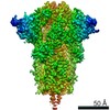 22352MC  7jjjC M: map data used to model this data C: citing same article ( |
|---|---|
| Similar structure data |


-Links
 PDBj
PDBj


- Assembly
| Deposited unit | 
|
|---|---|
| 1 |
|
-Components
| #1: Protein | Mass: 141176.125 Da / Num. of mol.: 3 / Mutation: R682Q, R683Q, R685Q, K986P, V987P Source method: isolated from a genetically manipulated source Source: (gene. exp.) Severe acute respiratory syndrome coronavirus 2Gene: S, 2 / Cell (production host): sf9 / Production host:   Spodoptera frugiperda (fall armyworm) / References: UniProt: P0DTC2 Spodoptera frugiperda (fall armyworm) / References: UniProt: P0DTC2#2: Polysaccharide | 2-acetamido-2-deoxy-beta-D-glucopyranose-(1-4)-2-acetamido-2-deoxy-beta-D-glucopyranose Source method: isolated from a genetically manipulated source #3: Sugar |   Type: D-saccharide, beta linking / Mass: 221.208 Da / Num. of mol.: 3 / Source method: obtained synthetically / Formula: C8H15NO6 Type: D-saccharide, beta linking / Mass: 221.208 Da / Num. of mol.: 3 / Source method: obtained synthetically / Formula: C8H15NO6#4: Chemical |   Mass: 604.813 Da / Num. of mol.: 3 / Source method: obtained synthetically / Formula: C32H60O10 / Feature type: SUBJECT OF INVESTIGATION Mass: 604.813 Da / Num. of mol.: 3 / Source method: obtained synthetically / Formula: C32H60O10 / Feature type: SUBJECT OF INVESTIGATION#5: Chemical |   Mass: 280.445 Da / Num. of mol.: 3 / Source method: obtained synthetically / Formula: C18H32O2 / Feature type: SUBJECT OF INVESTIGATION Mass: 280.445 Da / Num. of mol.: 3 / Source method: obtained synthetically / Formula: C18H32O2 / Feature type: SUBJECT OF INVESTIGATIONHas ligand of interest | Y | Has protein modification | Y | |
|---|
-Experimental details
-Experiment
| Experiment | Method: ELECTRON MICROSCOPY |
|---|---|
| EM experiment | Aggregation state: PARTICLE / 3D reconstruction method: single particle reconstruction |
- Sample preparation
| Component | Name: SARS-CoV-2 full-length spike / Type: COMPLEX / Entity ID: #1 / Source: RECOMBINANT | ||||||||||||||||||||
|---|---|---|---|---|---|---|---|---|---|---|---|---|---|---|---|---|---|---|---|---|---|
| Molecular weight | Experimental value: NO | ||||||||||||||||||||
| Source (natural) | Organism: Severe acute respiratory syndrome coronavirus 2 | ||||||||||||||||||||
| Source (recombinant) | Organism: Spodoptera frugiperda (fall armyworm) | ||||||||||||||||||||
| Buffer solution | pH: 7.2 | ||||||||||||||||||||
| Buffer component |
| ||||||||||||||||||||
| Specimen | Conc.: 0.4 mg/ml / Embedding applied: NO / Shadowing applied: NO / Staining applied: NO / Vitrification applied: YES | ||||||||||||||||||||
| Specimen support | Grid material: GOLD / Grid mesh size: 300 divisions/in. / Grid type: UltrAuFoil R1.2/1.3 | ||||||||||||||||||||
| Vitrification | Instrument: FEI VITROBOT MARK IV / Cryogen name: ETHANE / Humidity: 100 % / Chamber temperature: 283 K |
- Electron microscopy imaging
| Experimental equipment |  Model: Talos Arctica / Image courtesy: FEI Company |
|---|---|
| Microscopy | Model: FEI TALOS ARCTICA |
| Electron gun | Electron source:  FIELD EMISSION GUN / Accelerating voltage: 200 kV / Illumination mode: FLOOD BEAM FIELD EMISSION GUN / Accelerating voltage: 200 kV / Illumination mode: FLOOD BEAM |
| Electron lens | Mode: BRIGHT FIELD / Nominal magnification: 36000 X / Nominal defocus max: 1600 nm / Nominal defocus min: 500 nm / Cs: 2.7 mm / C2 aperture diameter: 70 µm / Alignment procedure: COMA FREE |
| Specimen holder | Cryogen: NITROGEN / Specimen holder model: FEI TITAN KRIOS AUTOGRID HOLDER |
| Image recording | Average exposure time: 11.5 sec. / Electron dose: 50 e/Å2 / Detector mode: COUNTING / Film or detector model: GATAN K2 SUMMIT (4k x 4k) / Num. of real images: 5506 |
| Image scans | Movie frames/image: 46 |
- Processing
| EM software |
| ||||||||||||||||||||||||||||||||||||||||
|---|---|---|---|---|---|---|---|---|---|---|---|---|---|---|---|---|---|---|---|---|---|---|---|---|---|---|---|---|---|---|---|---|---|---|---|---|---|---|---|---|---|
| CTF correction | Type: PHASE FLIPPING AND AMPLITUDE CORRECTION | ||||||||||||||||||||||||||||||||||||||||
| Symmetry | Point symmetry: C3 (3 fold cyclic) | ||||||||||||||||||||||||||||||||||||||||
| 3D reconstruction | Resolution: 3.6 Å / Resolution method: FSC 0.143 CUT-OFF / Num. of particles: 45374 / Algorithm: BACK PROJECTION / Symmetry type: POINT | ||||||||||||||||||||||||||||||||||||||||
| Atomic model building | Protocol: RIGID BODY FIT / Space: REAL | ||||||||||||||||||||||||||||||||||||||||
| Atomic model building | PDB-ID: 6VXX Accession code: 6VXX / Source name: PDB / Type: experimental model |