Biotechnology and Biological Sciences Research Council (BBSRC)
BB/P000940/1
United Kingdom
Wellcome Trust
202904/Z/16/Z
United Kingdom
Wellcome Trust
206181/Z/17/Z
United Kingdom
Biotechnology and Biological Sciences Research Council (BBSRC)
BB/L01386X/1
United Kingdom
Wellcome Trust
210701/Z/18/Z
United Kingdom
Wellcome Trust
106115/Z/14/Z
United Kingdom
Biotechnology and Biological Sciences Research Council (BBSRC)
BB/R000484/1
United Kingdom
Citation
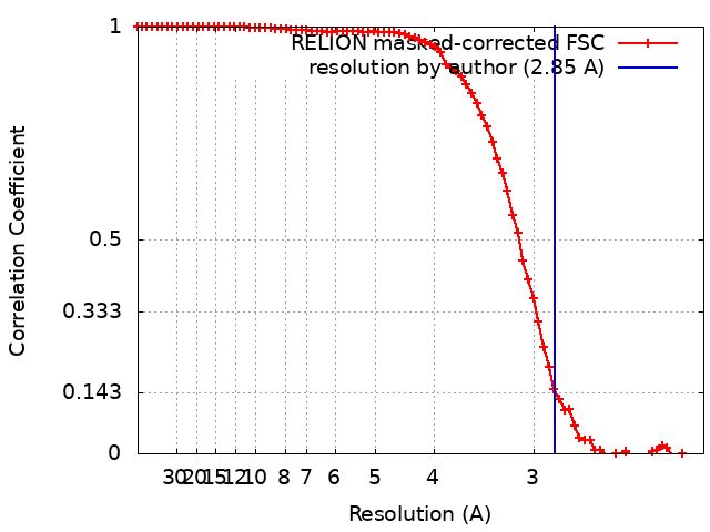
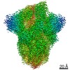
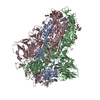
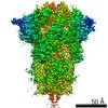
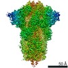
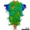
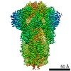
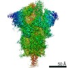
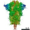
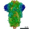
Journal: Science / Year: 2020 Title: Free fatty acid binding pocket in the locked structure of SARS-CoV-2 spike protein. Authors: Christine Toelzer / Kapil Gupta / Sathish K N Yadav / Ufuk Borucu / Andrew D Davidson / Maia Kavanagh Williamson / Deborah K Shoemark / Frederic Garzoni / Oskar Staufer / Rachel Milligan / ...Authors: Christine Toelzer / Kapil Gupta / Sathish K N Yadav / Ufuk Borucu / Andrew D Davidson / Maia Kavanagh Williamson / Deborah K Shoemark / Frederic Garzoni / Oskar Staufer / Rachel Milligan / Julien Capin / Adrian J Mulholland / Joachim Spatz / Daniel Fitzgerald / Imre Berger / Christiane Schaffitzel / Abstract: Coronavirus disease 2019 (COVID-19), caused by severe acute respiratory syndrome coronavirus 2 (SARS-CoV-2), represents a global crisis. Key to SARS-CoV-2 therapeutic development is unraveling the ...Coronavirus disease 2019 (COVID-19), caused by severe acute respiratory syndrome coronavirus 2 (SARS-CoV-2), represents a global crisis. Key to SARS-CoV-2 therapeutic development is unraveling the mechanisms that drive high infectivity, broad tissue tropism, and severe pathology. Our 2.85-angstrom cryo-electron microscopy structure of SARS-CoV-2 spike (S) glycoprotein reveals that the receptor binding domains tightly bind the essential free fatty acid linoleic acid (LA) in three composite binding pockets. A similar pocket also appears to be present in the highly pathogenic severe acute respiratory syndrome coronavirus (SARS-CoV) and Middle East respiratory syndrome coronavirus (MERS-CoV). LA binding stabilizes a locked S conformation, resulting in reduced angiotensin-converting enzyme 2 (ACE2) interaction in vitro In human cells, LA supplementation synergizes with the COVID-19 drug remdesivir, suppressing SARS-CoV-2 replication. Our structure directly links LA and S, setting the stage for intervention strategies that target LA binding by SARS-CoV-2.
History
Deposition
Jun 7, 2020
-
Header (metadata) release
Sep 30, 2020
-
Map release
Sep 30, 2020
-
Update
Nov 13, 2024
-
Current status
Nov 13, 2024
Processing site: PDBe / Status: Released
-
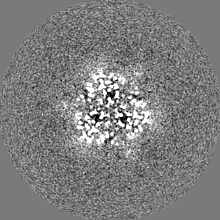
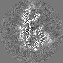
Structure visualization
Movie
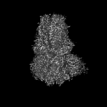
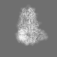
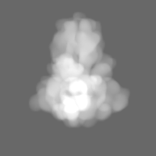
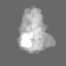
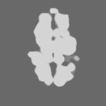
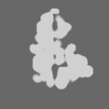
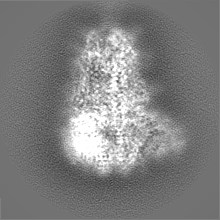
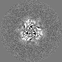
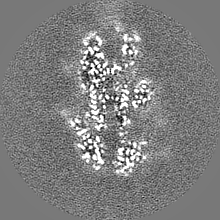
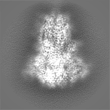
Surface view with section colored by density value
Film or detector model: GATAN K2 SUMMIT (4k x 4k) / Detector mode: SUPER-RESOLUTION / Number grids imaged: 1 / Number real images: 3289 / Average exposure time: 11.0 sec. / Average electron dose: 60.5 e/Å2
Electron beam
Acceleration voltage: 200 kV / Electron source: FIELD EMISSION GUN
In the structure databanks used in Yorodumi, some data are registered as the other names, "COVID-19 virus" and "2019-nCoV". Here are the details of the virus and the list of structure data.
Jan 31, 2019. EMDB accession codes are about to change! (news from PDBe EMDB page)
EMDB accession codes are about to change! (news from PDBe EMDB page)
The allocation of 4 digits for EMDB accession codes will soon come to an end. Whilst these codes will remain in use, new EMDB accession codes will include an additional digit and will expand incrementally as the available range of codes is exhausted. The current 4-digit format prefixed with “EMD-” (i.e. EMD-XXXX) will advance to a 5-digit format (i.e. EMD-XXXXX), and so on. It is currently estimated that the 4-digit codes will be depleted around Spring 2019, at which point the 5-digit format will come into force.
The EM Navigator/Yorodumi systems omit the EMD- prefix.
Related info.:Q: What is EMD? / ID/Accession-code notation in Yorodumi/EM Navigator
Yorodumi is a browser for structure data from EMDB, PDB, SASBDB, etc.
This page is also the successor to EM Navigator detail page, and also detail information page/front-end page for Omokage search.
The word "yorodu" (or yorozu) is an old Japanese word meaning "ten thousand". "mi" (miru) is to see.
Related info.:EMDB / PDB / SASBDB / Comparison of 3 databanks / Yorodumi Search / Aug 31, 2016. New EM Navigator & Yorodumi / Yorodumi Papers / Jmol/JSmol / Function and homology information / Changes in new EM Navigator and Yorodumi
 Movie
Movie Controller


 Open data
 Basic information
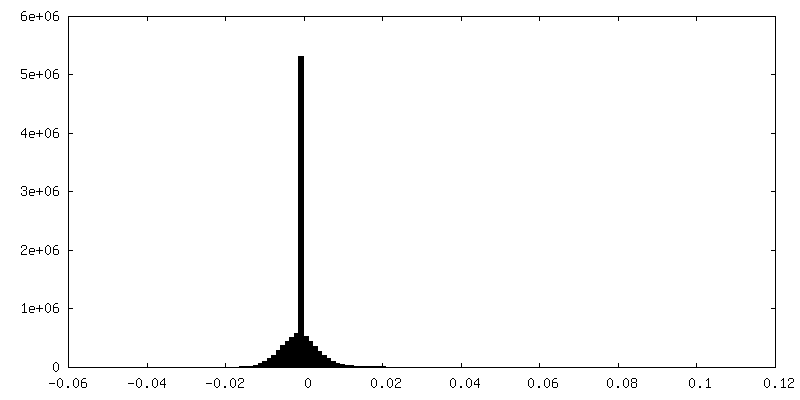
 Map data
 Sample
 Keywords
 Function and homology information
 Severe acute respiratory syndrome coronavirus 2
 Authors
 United Kingdom, 7 items
  Citation

 Structure visualization
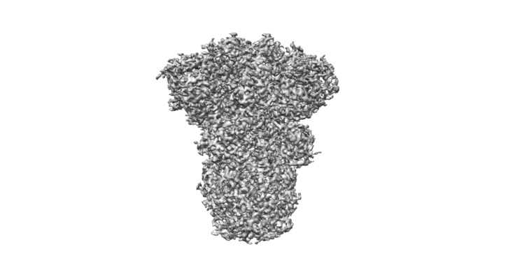
 Downloads & links
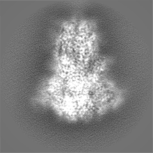
 emd_11145.png
 http://ftp.pdbj.org/pub/emdb/structures/EMD-11145


 Z (Sec.)
 Y (Row.)
 X (Col.)


 Sample components
 Trichoplusia ni (cabbage looper)

 Processing
 Electron microscopy
 FIELD EMISSION GUN