 Movie
Movie Controller


+ Open data

- Basic information
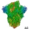
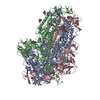
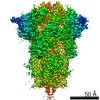
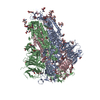
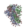
| Entry | Database: PDB / ID: 6zb5 | ||||||||||||||||||||||||
|---|---|---|---|---|---|---|---|---|---|---|---|---|---|---|---|---|---|---|---|---|---|---|---|---|---|
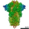
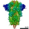
| Title | SARS CoV-2 Spike protein, Closed conformation, C3 symmetry | ||||||||||||||||||||||||
 Components Components | Spike glycoprotein | ||||||||||||||||||||||||
 Keywords Keywords | VIRAL PROTEIN / SARS-Cov2 / COVID / Linoleic Acid / Spike | ||||||||||||||||||||||||
| Function / homology |  Function and homology information Function and homology informationsymbiont-mediated disruption of host tissue / Maturation of spike protein / Translation of Structural Proteins / Virion Assembly and Release / host cell surface / host extracellular region / symbiont-mediated-mediated suppression of host tetherin activity / Induction of Cell-Cell Fusion / structural constituent of virion / positive regulation of viral entry into host cell ...symbiont-mediated disruption of host tissue / Maturation of spike protein / Translation of Structural Proteins / Virion Assembly and Release / host cell surface / host extracellular region / symbiont-mediated-mediated suppression of host tetherin activity / Induction of Cell-Cell Fusion / structural constituent of virion / positive regulation of viral entry into host cell / membrane fusion / host cell endoplasmic reticulum-Golgi intermediate compartment membrane / Attachment and Entry / entry receptor-mediated virion attachment to host cell / receptor-mediated virion attachment to host cell / host cell surface receptor binding / symbiont-mediated suppression of host innate immune response / endocytosis involved in viral entry into host cell / receptor ligand activity / fusion of virus membrane with host plasma membrane / fusion of virus membrane with host endosome membrane / viral envelope / symbiont entry into host cell / virion attachment to host cell / host cell plasma membrane / SARS-CoV-2 activates/modulates innate and adaptive immune responses / virion membrane / membrane / identical protein binding / plasma membrane Similarity search - Function | ||||||||||||||||||||||||
| Biological species |   Severe acute respiratory syndrome coronavirus 2 Severe acute respiratory syndrome coronavirus 2 | ||||||||||||||||||||||||
| Method | ELECTRON MICROSCOPY / single particle reconstruction / cryo EM / Resolution: 2.85 Å | ||||||||||||||||||||||||
 Authors Authors | Toelzer, C. / Gupta, K. / Yadav, S.K.N. / Burucu, U. / Schaffitzel, C. / Berger, I. | ||||||||||||||||||||||||
| Funding support |  United Kingdom, 7items United Kingdom, 7items
| ||||||||||||||||||||||||
 Citation Citation | Journal: Science / Year: 2020 Title: Free fatty acid binding pocket in the locked structure of SARS-CoV-2 spike protein. Authors: Christine Toelzer / Kapil Gupta / Sathish K N Yadav / Ufuk Borucu / Andrew D Davidson / Maia Kavanagh Williamson / Deborah K Shoemark / Frederic Garzoni / Oskar Staufer / Rachel Milligan / ...Authors: Christine Toelzer / Kapil Gupta / Sathish K N Yadav / Ufuk Borucu / Andrew D Davidson / Maia Kavanagh Williamson / Deborah K Shoemark / Frederic Garzoni / Oskar Staufer / Rachel Milligan / Julien Capin / Adrian J Mulholland / Joachim Spatz / Daniel Fitzgerald / Imre Berger / Christiane Schaffitzel /   Abstract: Coronavirus disease 2019 (COVID-19), caused by severe acute respiratory syndrome coronavirus 2 (SARS-CoV-2), represents a global crisis. Key to SARS-CoV-2 therapeutic development is unraveling the ...Coronavirus disease 2019 (COVID-19), caused by severe acute respiratory syndrome coronavirus 2 (SARS-CoV-2), represents a global crisis. Key to SARS-CoV-2 therapeutic development is unraveling the mechanisms that drive high infectivity, broad tissue tropism, and severe pathology. Our 2.85-angstrom cryo-electron microscopy structure of SARS-CoV-2 spike (S) glycoprotein reveals that the receptor binding domains tightly bind the essential free fatty acid linoleic acid (LA) in three composite binding pockets. A similar pocket also appears to be present in the highly pathogenic severe acute respiratory syndrome coronavirus (SARS-CoV) and Middle East respiratory syndrome coronavirus (MERS-CoV). LA binding stabilizes a locked S conformation, resulting in reduced angiotensin-converting enzyme 2 (ACE2) interaction in vitro In human cells, LA supplementation synergizes with the COVID-19 drug remdesivir, suppressing SARS-CoV-2 replication. Our structure directly links LA and S, setting the stage for intervention strategies that target LA binding by SARS-CoV-2. | ||||||||||||||||||||||||
| History |
|
- Structure visualization
| Movie |
Movie viewer |
|---|---|
| Structure viewer | Molecule: MolmilJmol/JSmol |
- Downloads & links
-Download
| PDBx/mmCIF format | 6zb5.cif.gz | 558.8 KB | Display | PDBx/mmCIF format |
|---|---|---|---|---|
| PDB format | pdb6zb5.ent.gz | 446 KB | Display | PDB format |
| PDBx/mmJSON format | 6zb5.json.gz | Tree view | PDBx/mmJSON format | |
| Others |  Other downloads Other downloads |
-Validation report
| Arichive directory | https://data.pdbj.org/pub/pdb/validation_reports/zb/6zb5ftp://data.pdbj.org/pub/pdb/validation_reports/zb/6zb5 | HTTPS FTP |
|---|
-Related structure data
| Related structure data | 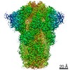 11145MC  6zb4C M: map data used to model this data C: citing same article ( |
|---|---|
| Similar structure data |


-Links
 PDBj
PDBj


- Assembly
| Deposited unit | 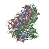
|
|---|---|
| 1 |
|
-Components
| #1: Protein | Mass: 139735.922 Da / Num. of mol.: 3 Source method: isolated from a genetically manipulated source Source: (gene. exp.) Severe acute respiratory syndrome coronavirus 2Gene: S, 2 / Production host:  Trichoplusia ni (cabbage looper) / References: UniProt: P0DTC2 Trichoplusia ni (cabbage looper) / References: UniProt: P0DTC2#2: Polysaccharide | #3: Sugar | ChemComp-NAG /   Type: D-saccharide, beta linking / Mass: 221.208 Da / Num. of mol.: 20 Type: D-saccharide, beta linking / Mass: 221.208 Da / Num. of mol.: 20Source method: isolated from a genetically manipulated source Formula: C8H15NO6 #4: Chemical |   Mass: 280.445 Da / Num. of mol.: 3 / Source method: obtained synthetically / Formula: C18H32O2 / Feature type: SUBJECT OF INVESTIGATION Mass: 280.445 Da / Num. of mol.: 3 / Source method: obtained synthetically / Formula: C18H32O2 / Feature type: SUBJECT OF INVESTIGATIONHas ligand of interest | Y | Has protein modification | Y | |
|---|
-Experimental details
-Experiment
| Experiment | Method: ELECTRON MICROSCOPY |
|---|---|
| EM experiment | Aggregation state: PARTICLE / 3D reconstruction method: single particle reconstruction |
- Sample preparation
| Component | Name: SARS CoV-2 Spike protein, Closed conformation, C3 symmetry Type: COMPLEX / Entity ID: #1 / Source: RECOMBINANT |
|---|---|
| Molecular weight | Value: 0.54 MDa / Experimental value: NO |
| Source (natural) | Organism: Severe acute respiratory syndrome coronavirus 2 |
| Source (recombinant) | Organism: Trichoplusia ni (cabbage looper) |
| Buffer solution | pH: 7.5 |
| Specimen | Conc.: 1.2 mg/ml / Embedding applied: NO / Shadowing applied: NO / Staining applied: NO / Vitrification applied: YES |
| Specimen support | Grid material: COPPER / Grid mesh size: 300 divisions/in. / Grid type: Quantifoil R1.2/1.3 |
| Vitrification | Cryogen name: ETHANE-PROPANE |
- Electron microscopy imaging
| Microscopy | Model: TFS TALOS |
|---|---|
| Electron gun | Electron source:  FIELD EMISSION GUN / Accelerating voltage: 200 kV / Illumination mode: FLOOD BEAM FIELD EMISSION GUN / Accelerating voltage: 200 kV / Illumination mode: FLOOD BEAM |
| Electron lens | Mode: BRIGHT FIELD / Nominal magnification: 130000 X / Nominal defocus max: 2000 nm / Nominal defocus min: 800 nm / Cs: 2.7 mm / C2 aperture diameter: 50 µm / Alignment procedure: COMA FREE |
| Specimen holder | Cryogen: NITROGEN / Specimen holder model: FEI TITAN KRIOS AUTOGRID HOLDER |
| Image recording | Average exposure time: 11 sec. / Electron dose: 60.5 e/Å2 / Detector mode: SUPER-RESOLUTION / Film or detector model: GATAN K2 SUMMIT (4k x 4k) / Num. of grids imaged: 1 / Num. of real images: 3289 |
| Image scans | Movie frames/image: 55 |
- Processing
| Software | Name: PHENIX / Version: 1.17_3644: / Classification: refinement | ||||||||||||||||||||||||
|---|---|---|---|---|---|---|---|---|---|---|---|---|---|---|---|---|---|---|---|---|---|---|---|---|---|
| EM software |
| ||||||||||||||||||||||||
| CTF correction | Type: PHASE FLIPPING AND AMPLITUDE CORRECTION | ||||||||||||||||||||||||
| Symmetry | Point symmetry: C3 (3 fold cyclic) | ||||||||||||||||||||||||
| 3D reconstruction | Resolution: 2.85 Å / Resolution method: FSC 0.143 CUT-OFF / Num. of particles: 217815 / Algorithm: FOURIER SPACE / Num. of class averages: 1 / Symmetry type: POINT | ||||||||||||||||||||||||
| Refinement | Cross valid method: NONE | ||||||||||||||||||||||||
| Refine LS restraints |
|
