 ムービー
ムービー コントローラー


+ データを開く

- 基本情報
基本情報
| 登録情報 | データベース: EMDB / ID: EMD-10641 | |||||||||
|---|---|---|---|---|---|---|---|---|---|---|
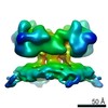
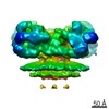
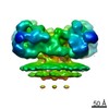
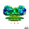
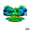
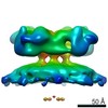
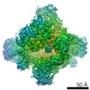
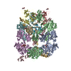
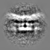
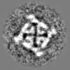
| タイトル | Structure of ryanodine receptor 1 in native membrane in the presence of DDM and EDTA | |||||||||
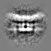
 マップデータ マップデータ | Structure of rabbit ryanodine receptor 1 in native membrane in the presence of DDM and EDTA | |||||||||
 試料 試料 |
| |||||||||
| 生物種 |  | |||||||||
| 手法 | サブトモグラム平均法 / クライオ電子顕微鏡法 / 解像度: 25.7 Å | |||||||||
 データ登録者 データ登録者 | Chen W / Kudryashev M | |||||||||
| 資金援助 |  ドイツ, 1件 ドイツ, 1件
| |||||||||
 引用 引用 | ジャーナル: EMBO Rep / 年: 2020 タイトル: Structure of RyR1 in native membranes. 著者: Wenbo Chen / Mikhail Kudryashev / 要旨: Ryanodine receptor 1 (RyR1) mediates excitation-contraction coupling by releasing Ca from sarcoplasmic reticulum (SR) to the cytoplasm of skeletal muscle cells. RyR1 activation is regulated by ...Ryanodine receptor 1 (RyR1) mediates excitation-contraction coupling by releasing Ca from sarcoplasmic reticulum (SR) to the cytoplasm of skeletal muscle cells. RyR1 activation is regulated by several proteins from both the cytoplasm and lumen of the SR. Here, we report the structure of RyR1 from native SR membranes in closed and open states. Compared to the previously reported structures of purified RyR1, our structure reveals helix-like densities traversing the bilayer approximately 5 nm from the RyR1 transmembrane domain and sarcoplasmic extensions linking RyR1 to a putative calsequestrin network. We document the primary conformation of RyR1 in situ and its structural variations. The activation of RyR1 is associated with changes in membrane curvature and movement in the sarcoplasmic extensions. Our results provide structural insight into the mechanism of RyR1 in its native environment. | |||||||||
| 履歴 |
|
- 構造の表示
構造の表示
| ムービー |
 ムービービューア ムービービューア |
|---|---|
| 構造ビューア | EMマップ: SurfViewMolmilJmol/JSmol |
| 添付画像 |

- ダウンロードとリンク
-EMDBアーカイブ
| マップデータ | emd_10641.map.gz | 3.6 MB | EMDBマップデータ形式 | |
|---|---|---|---|---|
| ヘッダ (付随情報) | emd-10641-v30.xmlemd-10641.xml | 14.1 KB 14.1 KB | 表示 表示 | EMDBヘッダ |
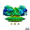
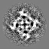
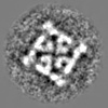
| 画像 | 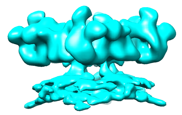 emd_10641.png emd_10641.png | 130.5 KB | ||
| マスクデータ | emd_10641_msk_1.map | 3.8 MB | マスクマップ | |
| その他 | emd_10641_half_map_1.map.gzemd_10641_half_map_2.map.gz | 3.6 MB 3.6 MB | ||
| アーカイブディレクトリ |  http://ftp.pdbj.org/pub/emdb/structures/EMD-10641ftp://ftp.pdbj.org/pub/emdb/structures/EMD-10641 http://ftp.pdbj.org/pub/emdb/structures/EMD-10641ftp://ftp.pdbj.org/pub/emdb/structures/EMD-10641 | HTTPS FTP |
-検証レポート
| 文書・要旨 | emd_10641_validation.pdf.gz | 357 KB | 表示 | EMDB検証レポート |
|---|---|---|---|---|
| 文書・詳細版 | emd_10641_full_validation.pdf.gz | 356.1 KB | 表示 | |
| XML形式データ | emd_10641_validation.xml.gz | 7.3 KB | 表示 | |
| アーカイブディレクトリ | https://ftp.pdbj.org/pub/emdb/validation_reports/EMD-10641ftp://ftp.pdbj.org/pub/emdb/validation_reports/EMD-10641 | HTTPS FTP |
-関連構造データ
| 関連構造データ | C: 同じ文献を引用 ( |
|---|---|
| 類似構造データ | |
| 電子顕微鏡画像生データ | EMPIAR-10349 (タイトル: Cryo Electron Tomograms of Membrane Fractions of Rabbit Skeletal Muscle for Structural Determination of RyR1 in SR Vesicles Data size: 254.2 Data #1: 82 tilt series (frames aligned my motioncorr2) in .st stacks (mrc format for imod) including .tlt, .xf and .defocus files [tilt series]) |

-リンク
| EMDBのページ | EMDB (EBI/PDBe) / EMDataResource |
|---|
-マップ
| ファイル | ダウンロード / ファイル: emd_10641.map.gz / 形式: CCP4 / 大きさ: 3.8 MB / タイプ: IMAGE STORED AS FLOATING POINT NUMBER (4 BYTES) | ||||||||||||||||||||||||||||||||||||||||||||||||||||||||||||
|---|---|---|---|---|---|---|---|---|---|---|---|---|---|---|---|---|---|---|---|---|---|---|---|---|---|---|---|---|---|---|---|---|---|---|---|---|---|---|---|---|---|---|---|---|---|---|---|---|---|---|---|---|---|---|---|---|---|---|---|---|---|
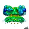
| 注釈 | Structure of rabbit ryanodine receptor 1 in native membrane in the presence of DDM and EDTA | ||||||||||||||||||||||||||||||||||||||||||||||||||||||||||||
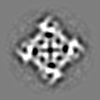
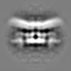
| 投影像・断面図 | 画像のコントロール
画像は Spider により作成 | ||||||||||||||||||||||||||||||||||||||||||||||||||||||||||||
| ボクセルのサイズ | X=Y=Z: 2.7 Å | ||||||||||||||||||||||||||||||||||||||||||||||||||||||||||||
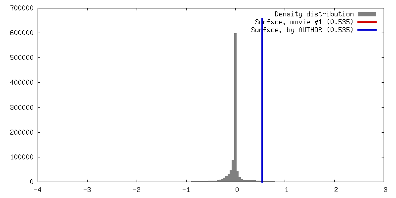
| 密度 |
| ||||||||||||||||||||||||||||||||||||||||||||||||||||||||||||
| 対称性 | 空間群: 1 | ||||||||||||||||||||||||||||||||||||||||||||||||||||||||||||
| 詳細 | EMDB XML:
CCP4マップ ヘッダ情報:
| ||||||||||||||||||||||||||||||||||||||||||||||||||||||||||||
 Z (Sec.)
 Y (Row.)
 X (Col.)


-添付データ
-マスク #1
| ファイル | emd_10641_msk_1.map | ||||||||||||
|---|---|---|---|---|---|---|---|---|---|---|---|---|---|
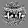
| 投影像・断面図 |
| ||||||||||||
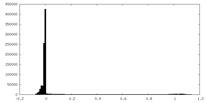
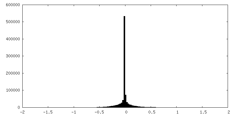
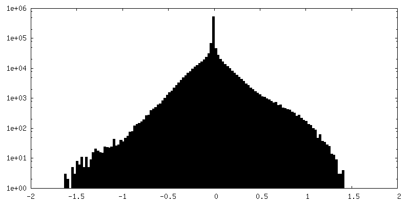
| 密度ヒストグラム |


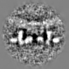
-ハーフマップ: Structure of rabbit ryanodine receptor 1 in native...
| ファイル | emd_10641_half_map_1.map | ||||||||||||
|---|---|---|---|---|---|---|---|---|---|---|---|---|---|
| 注釈 | Structure of rabbit ryanodine receptor 1 in native membrane in the presence of DDM and EDTA (half map 2) | ||||||||||||
| 投影像・断面図 |
| ||||||||||||
| 密度ヒストグラム |

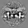

-ハーフマップ: Structure of rabbit ryanodine receptor 1 in native...
| ファイル | emd_10641_half_map_2.map | ||||||||||||
|---|---|---|---|---|---|---|---|---|---|---|---|---|---|
| 注釈 | Structure of rabbit ryanodine receptor 1 in native membrane in the presence of DDM and EDTA (half map 1) | ||||||||||||
| 投影像・断面図 |
| ||||||||||||
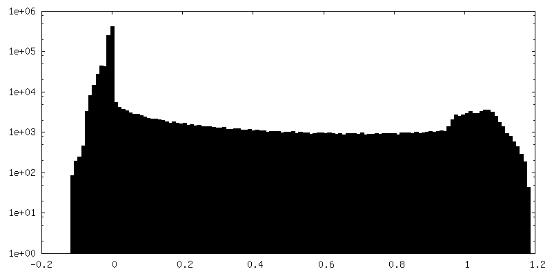
| 密度ヒストグラム |


- 試料の構成要素
-全体 : Rabbit ryanodine receptor 1 in native membrane in the presence of...
| 全体 | 名称: Rabbit ryanodine receptor 1 in native membrane in the presence of DDM and EDTA |
|---|---|
| 要素 |
|
-超分子 #1: Rabbit ryanodine receptor 1 in native membrane in the presence of...
| 超分子 | 名称: Rabbit ryanodine receptor 1 in native membrane in the presence of DDM and EDTA タイプ: complex / ID: 1 / 親要素: 0 |
|---|---|
| 由来(天然) | 生物種: |
-実験情報
-構造解析
| 手法 | クライオ電子顕微鏡法 |
|---|---|
 解析 解析 | サブトモグラム平均法 |
| 試料の集合状態 | tissue |
-試料調製
| 緩衝液 | pH: 7.1 |
|---|---|
| 凍結 | 凍結剤: ETHANE |
- 電子顕微鏡法
| 顕微鏡 | FEI TITAN KRIOS |
|---|---|
| 撮影 | フィルム・検出器のモデル: GATAN K2 SUMMIT (4k x 4k) 検出モード: COUNTING / 平均電子線量: 2.0 e/Å2 |
| 電子線 | 加速電圧: 300 kV / 電子線源:  FIELD EMISSION GUN FIELD EMISSION GUN |
| 電子光学系 | 照射モード: FLOOD BEAM / 撮影モード: BRIGHT FIELD |
| 実験機器 |  モデル: Titan Krios / 画像提供: FEI Company |
