 Movie
Movie Controller

[English] 日本語
 Yorodumi
- PDB-6r7f: Structural basis of Cullin-2 RING E3 ligase regulation by the COP... -
+ Open data

- Basic information
| Entry | Database: PDB / ID: 6r7f | ||||||
|---|---|---|---|---|---|---|---|
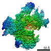
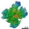
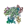
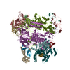
| Title | Structural basis of Cullin-2 RING E3 ligase regulation by the COP9 signalosome | ||||||
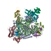
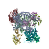
 Components Components |
| ||||||
 Keywords Keywords | LIGASE / Cullin-Ring E3 Ligase COP9 signalosome neddylation | ||||||
| Function / homology |  Function and homology information Function and homology informationCOP9 signalosome assembly / trophectodermal cell proliferation / macrophage migration inhibitory factor binding / regulation of IRE1-mediated unfolded protein response / exosomal secretion / GTPase inhibitor activity / deNEDDylase activity / protein deneddylation / regulation of protein neddylation / activation of NF-kappaB-inducing kinase activity ...COP9 signalosome assembly / trophectodermal cell proliferation / macrophage migration inhibitory factor binding / regulation of IRE1-mediated unfolded protein response / exosomal secretion / GTPase inhibitor activity / deNEDDylase activity / protein deneddylation / regulation of protein neddylation / activation of NF-kappaB-inducing kinase activity / eukaryotic translation initiation factor 3 complex / regulation of cellular response to hypoxia / negative regulation of beige fat cell differentiation / negative regulation of receptor signaling pathway via JAK-STAT / RHOBTB3 ATPase cycle / cellular response to camptothecin / COP9 signalosome / cullin-RING-type E3 NEDD8 transferase / NEDD8 transferase activity / cullin-RING ubiquitin ligase complex / Cul7-RING ubiquitin ligase complex / cellular response to chemical stress / Loss of Function of FBXW7 in Cancer and NOTCH1 Signaling / target-directed miRNA degradation / elongin complex / positive regulation of protein autoubiquitination / RNA polymerase II transcription initiation surveillance / protein neddylation / Replication of the SARS-CoV-1 genome / transcription elongation factor activity / Hydrolases; Acting on peptide bonds (peptidases) / NEDD8 ligase activity / protein K27-linked ubiquitination / RHOBTB1 GTPase cycle / negative regulation of response to oxidative stress / regulation of JNK cascade / regulation of DNA damage response, signal transduction by p53 class mediator / VCB complex / inner cell mass cell proliferation / Cul5-RING ubiquitin ligase complex / metal-dependent deubiquitinase activity / ubiquitin-ubiquitin ligase activity / ubiquitin-dependent protein catabolic process via the C-end degron rule pathway / SCF ubiquitin ligase complex / Cul2-RING ubiquitin ligase complex / SUMOylation of ubiquitinylation proteins / Cul3-RING ubiquitin ligase complex / negative regulation of type I interferon production / SCF-dependent proteasomal ubiquitin-dependent protein catabolic process / Cul4A-RING E3 ubiquitin ligase complex / Prolactin receptor signaling / Cul4-RING E3 ubiquitin ligase complex / negative regulation of mitophagy / Cul4B-RING E3 ubiquitin ligase complex / ubiquitin ligase complex scaffold activity / TGF-beta receptor signaling activates SMADs / negative regulation of transcription elongation by RNA polymerase II / regulation of proteolysis / Pausing and recovery of Tat-mediated HIV elongation / Tat-mediated HIV elongation arrest and recovery / : / HIV elongation arrest and recovery / Pausing and recovery of HIV elongation / regulation of postsynapse assembly / response to light stimulus / skeletal muscle cell differentiation / cullin family protein binding / anatomical structure morphogenesis / negative regulation of signal transduction / protein monoubiquitination / Tat-mediated elongation of the HIV-1 transcript / Formation of HIV-1 elongation complex containing HIV-1 Tat / Formation of HIV elongation complex in the absence of HIV Tat / ubiquitin-like ligase-substrate adaptor activity / site of DNA damage / RNA Polymerase II Transcription Elongation / Formation of RNA Pol II elongation complex / JNK cascade / signal transduction in response to DNA damage / Nuclear events stimulated by ALK signaling in cancer / negative regulation of TORC1 signaling / protein K48-linked ubiquitination / transcription-coupled nucleotide-excision repair / negative regulation of insulin receptor signaling pathway / translation initiation factor activity / RNA Polymerase II Pre-transcription Events / regulation of cellular response to insulin stimulus / positive regulation of TORC1 signaling / post-translational protein modification / intrinsic apoptotic signaling pathway / ciliary tip / T cell activation / protein modification process / transcription corepressor binding / Regulation of BACH1 activity / negative regulation of autophagy / protein serine/threonine kinase binding / negative regulation of canonical NF-kappaB signal transduction / TP53 Regulates Transcription of DNA Repair Genes / positive regulation of cell differentiation Similarity search - Function | ||||||
| Biological species |  Homo sapiens (human) Homo sapiens (human) | ||||||
| Method | ELECTRON MICROSCOPY / single particle reconstruction / cryo EM / Resolution: 8.2 Å | ||||||
 Authors Authors | Faull, S.V. / Lau, A.M.C. / Martens, C. / Ahdash, Z. / Yebenes, H. / Schmidt, C. / Beuron, F. / Cronin, N.B. / Morris, E.P. / Politis, A. | ||||||
| Funding support |  United Kingdom, 1items United Kingdom, 1items
| ||||||
 Citation Citation | Journal: Nat Commun / Year: 2019 Title: Structural basis of Cullin 2 RING E3 ligase regulation by the COP9 signalosome. Authors: Sarah V Faull / Andy M C Lau / Chloe Martens / Zainab Ahdash / Kjetil Hansen / Hugo Yebenes / Carla Schmidt / Fabienne Beuron / Nora B Cronin / Edward P Morris / Argyris Politis /   Abstract: Cullin-Ring E3 Ligases (CRLs) regulate a multitude of cellular pathways through specific substrate receptors. The COP9 signalosome (CSN) deactivates CRLs by removing NEDD8 from activated Cullins. ...Cullin-Ring E3 Ligases (CRLs) regulate a multitude of cellular pathways through specific substrate receptors. The COP9 signalosome (CSN) deactivates CRLs by removing NEDD8 from activated Cullins. Here we present structures of the neddylated and deneddylated CSN-CRL2 complexes by combining single-particle cryo-electron microscopy (cryo-EM) with chemical cross-linking mass spectrometry (XL-MS). These structures suggest a conserved mechanism of CSN activation, consisting of conformational clamping of the CRL2 substrate by CSN2/CSN4, release of the catalytic CSN5/CSN6 heterodimer and finally activation of the CSN5 deneddylation machinery. Using hydrogen-deuterium exchange (HDX)-MS we show that CRL2 activates CSN5/CSN6 in a neddylation-independent manner. The presence of NEDD8 is required to activate the CSN5 active site. Overall, by synergising cryo-EM with MS, we identify sensory regions of the CSN that mediate its stepwise activation and provide a framework for understanding the regulatory mechanism of other Cullin family members. | ||||||
| History |
|
- Structure visualization
| Movie |
Movie viewer |
|---|---|
| Structure viewer | Molecule: MolmilJmol/JSmol |
- Downloads & links
-Download
| PDBx/mmCIF format | 6r7f.cif.gz | 720.3 KB | Display | PDBx/mmCIF format |
|---|---|---|---|---|
| PDB format | pdb6r7f.ent.gz | 558.5 KB | Display | PDB format |
| PDBx/mmJSON format | 6r7f.json.gz | Tree view | PDBx/mmJSON format | |
| Others |  Other downloads Other downloads |
-Validation report
| Arichive directory | https://data.pdbj.org/pub/pdb/validation_reports/r7/6r7fftp://data.pdbj.org/pub/pdb/validation_reports/r7/6r7f | HTTPS FTP |
|---|
-Related structure data
| Related structure data | 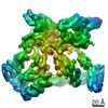 4739MC 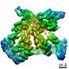 4736C 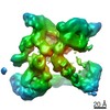 4741C 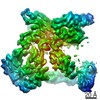 4742C 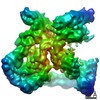 4744C 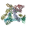 6r6hC 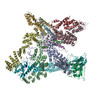 6r7hC  6r7iC 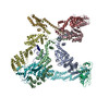 6r7nC M: map data used to model this data C: citing same article ( |
|---|---|
| Similar structure data |

-Links
 PDBj
PDBj


- Assembly
| Deposited unit | 
|
|---|---|
| 1 |
|
-Components
-COP9 signalosome complex subunit ... , 8 types, 8 molecules ABCDEFHG
| #1: Protein | Mass: 49253.535 Da / Num. of mol.: 1 Source method: isolated from a genetically manipulated source Source: (gene. exp.) Homo sapiens (human) / Gene: GPS1, COPS1, CSN1 / Production host:   Spodoptera frugiperda (fall armyworm) / References: UniProt: Q13098 Spodoptera frugiperda (fall armyworm) / References: UniProt: Q13098 |
|---|---|
| #2: Protein | Mass: 51664.570 Da / Num. of mol.: 1 Source method: isolated from a genetically manipulated source Source: (gene. exp.) Homo sapiens (human) / Gene: COPS2, CSN2, TRIP15 / Production host: Spodoptera frugiperda (fall armyworm) / References: UniProt: P61201 |
| #3: Protein | Mass: 45808.816 Da / Num. of mol.: 1 Source method: isolated from a genetically manipulated source Source: (gene. exp.) Homo sapiens (human) / Gene: COPS3, CSN3 / Production host: Spodoptera frugiperda (fall armyworm) / References: UniProt: Q9UNS2 |
| #4: Protein | Mass: 46322.688 Da / Num. of mol.: 1 Source method: isolated from a genetically manipulated source Source: (gene. exp.) Homo sapiens (human) / Gene: COPS4, CSN4 / Production host: Spodoptera frugiperda (fall armyworm) / References: UniProt: Q9BT78 |
| #5: Protein | Mass: 35184.059 Da / Num. of mol.: 1 Source method: isolated from a genetically manipulated source Source: (gene. exp.) Homo sapiens (human) / Gene: COPS5, CSN5, JAB1 / Production host: Spodoptera frugiperda (fall armyworm)References: UniProt: Q92905, Hydrolases; Acting on peptide bonds (peptidases) |
| #6: Protein | Mass: 32507.193 Da / Num. of mol.: 1 Source method: isolated from a genetically manipulated source Source: (gene. exp.) Homo sapiens (human) / Gene: COPS6, CSN6, HVIP / Production host: Spodoptera frugiperda (fall armyworm) / References: UniProt: Q7L5N1 |
| #7: Protein | Mass: 23245.543 Da / Num. of mol.: 1 Source method: isolated from a genetically manipulated source Source: (gene. exp.) Homo sapiens (human) / Gene: COPS8, CSN8 / Production host: Spodoptera frugiperda (fall armyworm) / References: UniProt: Q99627 |
| #8: Protein | Mass: 23429.709 Da / Num. of mol.: 1 Source method: isolated from a genetically manipulated source Source: (gene. exp.) Homo sapiens (human) / Gene: COPS7B, CSN7B / Production host: Spodoptera frugiperda (fall armyworm) / References: UniProt: Q9H9Q2 |
-Protein , 6 types, 6 molecules VPQORN
| #9: Protein | Mass: 18558.162 Da / Num. of mol.: 1 Source method: isolated from a genetically manipulated source Source: (gene. exp.) Homo sapiens (human) / Gene: VHL / Production host: Spodoptera frugiperda (fall armyworm) / References: UniProt: P40337 |
|---|---|
| #10: Protein | Mass: 13147.781 Da / Num. of mol.: 1 Source method: isolated from a genetically manipulated source Source: (gene. exp.) Homo sapiens (human) / Gene: ELOB, TCEB2 / Production host: Spodoptera frugiperda (fall armyworm) / References: UniProt: Q15370 |
| #11: Protein | Mass: 12485.135 Da / Num. of mol.: 1 Source method: isolated from a genetically manipulated source Source: (gene. exp.) Homo sapiens (human) / Gene: ELOC, TCEB1 / Production host: Spodoptera frugiperda (fall armyworm) / References: UniProt: Q15369 |
| #12: Protein | Mass: 87098.930 Da / Num. of mol.: 1 Source method: isolated from a genetically manipulated source Source: (gene. exp.) Homo sapiens (human) / Gene: CUL2 / Production host: Spodoptera frugiperda (fall armyworm) / References: UniProt: Q13617 |
| #13: Protein | Mass: 10655.229 Da / Num. of mol.: 1 Source method: isolated from a genetically manipulated source Source: (gene. exp.) Homo sapiens (human) / Production host: Spodoptera frugiperda (fall armyworm) / References: UniProt: P62877*PLUS |
| #14: Protein | Mass: 8573.978 Da / Num. of mol.: 1 Source method: isolated from a genetically manipulated source Source: (gene. exp.) Homo sapiens (human) / Gene: NEDD8 / Production host: Spodoptera frugiperda (fall armyworm) / References: UniProt: Q15843 |
-Experimental details
-Experiment
| Experiment | Method: ELECTRON MICROSCOPY |
|---|---|
| EM experiment | Aggregation state: PARTICLE / 3D reconstruction method: single particle reconstruction |
- Sample preparation
| Component | Name: Ternary complex of COP9 signalosome (CSN) and neddylated Cullin-Ring E3 ligase CRL (cullin-2 with Rbx1, Elongin-B, Elongin-C and VHL) Type: COMPLEX / Entity ID: all / Source: RECOMBINANT |
|---|---|
| Source (natural) | Organism: Homo sapiens (human) |
| Source (recombinant) | Organism: Spodoptera frugiperda (fall armyworm) |
| Buffer solution | pH: 7.5 Details: 15 mM HEPES pH 7.5 100 mM NaCL 0.5 mM DTT 1% Glycerol |
| Specimen | Embedding applied: NO / Shadowing applied: NO / Staining applied: NO / Vitrification applied: YES |
| Vitrification | Instrument: FEI VITROBOT MARK IV / Cryogen name: ETHANE |
- Electron microscopy imaging
| Experimental equipment |  Model: Titan Krios / Image courtesy: FEI Company |
|---|---|
| Microscopy | Model: FEI TITAN KRIOS |
| Electron gun | Electron source:  FIELD EMISSION GUN / Accelerating voltage: 300 kV / Illumination mode: FLOOD BEAM FIELD EMISSION GUN / Accelerating voltage: 300 kV / Illumination mode: FLOOD BEAM |
| Electron lens | Mode: BRIGHT FIELD / Nominal defocus max: 3000 nm / Nominal defocus min: 1800 nm / C2 aperture diameter: 100 µm |
| Specimen holder | Specimen holder model: FEI TITAN KRIOS AUTOGRID HOLDER |
| Image recording | Electron dose: 45 e/Å2 / Detector mode: COUNTING / Film or detector model: GATAN K2 QUANTUM (4k x 4k) |
- Processing
| CTF correction | Type: PHASE FLIPPING AND AMPLITUDE CORRECTION |
|---|---|
| 3D reconstruction | Resolution: 8.2 Å / Resolution method: FSC 0.143 CUT-OFF / Num. of particles: 20055 / Symmetry type: POINT |
