- EMDB-10533: Horse spleen apoferritin deposited with a microfluidic sprayer -
+
Open data
ID or keywords:
Loading...
-
Basic information
Entry
Database: EMDB / ID: EMD-10533
Title
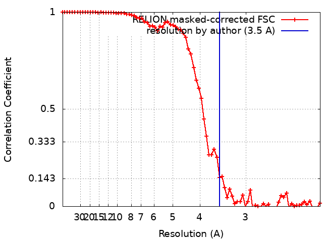
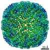
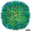
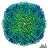
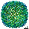
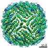
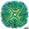
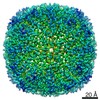
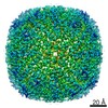
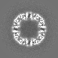
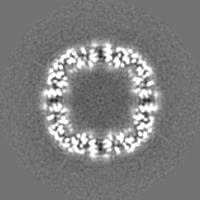
Horse spleen apoferritin deposited with a microfluidic sprayer
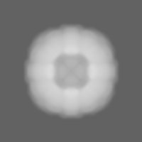
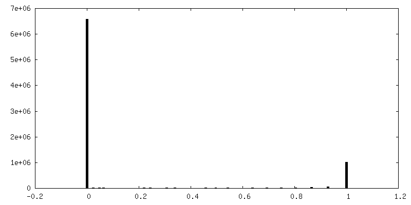
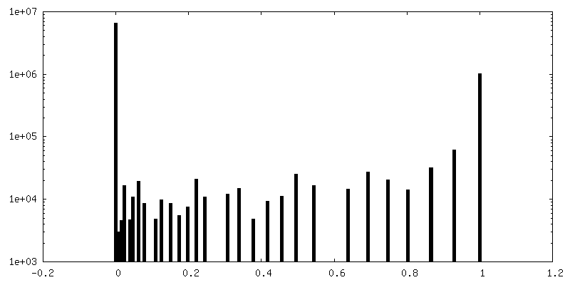
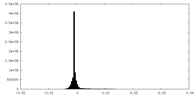
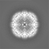
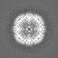
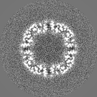
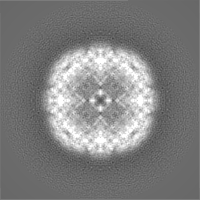
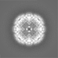
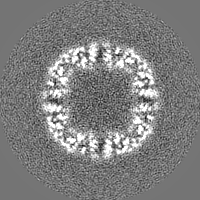
Map data
Final sharpened map
Sample
Complex: apoferritin from equine spleen
Function / homology
Function and homology information
ferritin complex / autolysosome / ferric iron binding / autophagosome / iron ion transport / ferrous iron binding / cytoplasmic vesicle / intracellular iron ion homeostasis / iron ion binding / cytoplasm Similarity search - Function
Biotechnology and Biological Sciences Research Council
BB/P026397/1
United Kingdom
Citation
Journal: Acta Crystallogr D Struct Biol / Year: 2020 Title: Sample deposition onto cryo-EM grids: from sprays to jets and back. Authors: David P Klebl / Diana C F Monteiro / Dimitrios Kontziampasis / Florian Kopf / Frank Sobott / Howard D White / Martin Trebbin / Stephen P Muench / Abstract: Despite the great strides made in the field of single-particle cryogenic electron microscopy (cryo-EM) in microscope design, direct electron detectors and new processing suites, the area of sample ...Despite the great strides made in the field of single-particle cryogenic electron microscopy (cryo-EM) in microscope design, direct electron detectors and new processing suites, the area of sample preparation is still far from ideal. Traditionally, sample preparation involves blotting, which has been used to achieve high resolution, particularly for well behaved samples such as apoferritin. However, this approach is flawed since the blotting process can have adverse effects on some proteins and protein complexes, and the long blot time increases exposure to the damaging air-water interface. To overcome these problems, new blotless approaches have been designed for the direct deposition of the sample on the grid. Here, different methods of producing droplets for sample deposition are compared. Using gas dynamic virtual nozzles, small and high-velocity droplets were deposited on cryo-EM grids, which spread sufficiently for high-resolution cryo-EM imaging. For those wishing to pursue a similar approach, an overview is given of the current use of spray technology for cryo-EM grid preparation and areas for enhancement are pointed out. It is further shown how the broad aspects of sprayer design and operation conditions can be utilized to improve grid quality reproducibly.
History
Deposition
Dec 8, 2019
-
Header (metadata) release
Apr 22, 2020
-
Map release
Apr 22, 2020
-
Update
Apr 22, 2020
-
Current status
Apr 22, 2020
Processing site: PDBe / Status: Released
-
Structure visualization
Movie
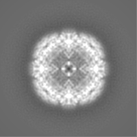
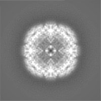
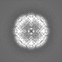
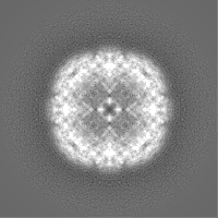
Surface view with section colored by density value
Model: Quantifoil R1.2/1.3 / Material: COPPER / Mesh: 300 / Support film - Material: CARBON / Support film - topology: HOLEY / Pretreatment - Type: GLOW DISCHARGE / Pretreatment - Atmosphere: AIR / Pretreatment - Pressure: 0.01 kPa
Vitrification
Cryogen name: ETHANE / Chamber humidity: 85 % / Chamber temperature: 295 K / Instrument: HOMEMADE PLUNGER Details: sample sprayed onto grid using microfluidic nozzle and frozen 36 ms after spraying.
-
Electron microscopy
Microscope
FEI TITAN KRIOS
Specialist optics
Energy filter - Name: GIF Bioquantum / Energy filter - Slit width: 20 eV
Image recording
Film or detector model: GATAN K2 SUMMIT (4k x 4k) / Detector mode: COUNTING / Number grids imaged: 1 / Number real images: 690 / Average exposure time: 8.0 sec. / Average electron dose: 60.0 e/Å2
Electron beam
Acceleration voltage: 300 kV / Electron source: FIELD EMISSION GUN
In the structure databanks used in Yorodumi, some data are registered as the other names, "COVID-19 virus" and "2019-nCoV". Here are the details of the virus and the list of structure data.
Jan 31, 2019. EMDB accession codes are about to change! (news from PDBe EMDB page)
EMDB accession codes are about to change! (news from PDBe EMDB page)
The allocation of 4 digits for EMDB accession codes will soon come to an end. Whilst these codes will remain in use, new EMDB accession codes will include an additional digit and will expand incrementally as the available range of codes is exhausted. The current 4-digit format prefixed with “EMD-” (i.e. EMD-XXXX) will advance to a 5-digit format (i.e. EMD-XXXXX), and so on. It is currently estimated that the 4-digit codes will be depleted around Spring 2019, at which point the 5-digit format will come into force.
The EM Navigator/Yorodumi systems omit the EMD- prefix.
Related info.:Q: What is EMD? / ID/Accession-code notation in Yorodumi/EM Navigator
Yorodumi is a browser for structure data from EMDB, PDB, SASBDB, etc.
This page is also the successor to EM Navigator detail page, and also detail information page/front-end page for Omokage search.
The word "yorodu" (or yorozu) is an old Japanese word meaning "ten thousand". "mi" (miru) is to see.
Related info.:EMDB / PDB / SASBDB / Comparison of 3 databanks / Yorodumi Search / Aug 31, 2016. New EM Navigator & Yorodumi / Yorodumi Papers / Jmol/JSmol / Function and homology information / Changes in new EM Navigator and Yorodumi
 Movie
Movie Controller


 Open data
 Basic information
 Map data
 Sample
 Function and homology information
 Authors
 United Kingdom, 1 items
  Citation

 Structure visualization
 Downloads & links
 emd_10533.png
 http://ftp.pdbj.org/pub/emdb/structures/EMD-10533
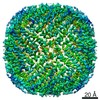


 Z (Sec.)
 Y (Row.)
 X (Col.)


 Sample components
 Processing
 Electron microscopy
 FIELD EMISSION GUN