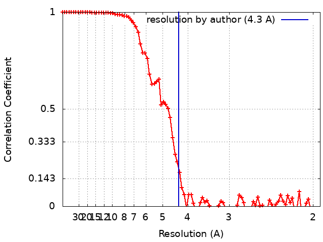
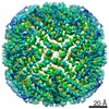
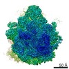
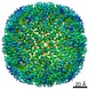
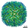
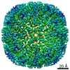
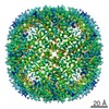
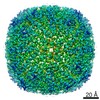
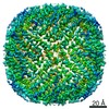
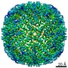
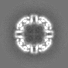
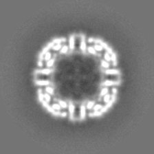
- EMDB-20157: Apoferritin from equine spleen at 4.3 Angstrom -
+
Open data
ID or keywords:
Loading...
-
Basic information
Entry
Database: EMDB / ID: EMD-20157
Title
Apoferritin from equine spleen at 4.3 Angstrom
Map data
Sharpen map
Sample
Complex: Apoferritin from equine spleen.
Function / homology
Function and homology information
ferritin complex / autolysosome / ferric iron binding / autophagosome / iron ion transport / ferrous iron binding / cytoplasmic vesicle / intracellular iron ion homeostasis / iron ion binding / cytoplasm Similarity search - Function
National Institutes of Health/National Human Genome Research Institute
R01GM108753
United States
Citation
Journal: IUCrJ / Year: 2019 Title: Throughput and resolution with a next-generation direct electron detector. Authors: Joshua H Mendez / Atousa Mehrani / Peter Randolph / Scott Stagg / Abstract: Direct electron detectors (DEDs) have revolutionized cryo-electron microscopy (cryo-EM) by facilitating the correction of beam-induced motion and radiation damage, and also by providing high- ...Direct electron detectors (DEDs) have revolutionized cryo-electron microscopy (cryo-EM) by facilitating the correction of beam-induced motion and radiation damage, and also by providing high-resolution image capture. A new-generation DED, the DE64, has been developed by Direct Electron that has good performance in both integrating and counting modes. The camera has been characterized in both modes in terms of image quality, throughput and resolution of cryo-EM reconstructions. The modulation transfer function, noise power spectrum and detective quantum efficiency (DQE) were determined for both modes, as well as the number of images per unit time. Although the DQE for counting mode was superior to that for integrating mode, the data-collection throughput for this mode was more than ten times slower. Since throughput and resolution are related in single-particle cryo-EM, data for apoferritin were collected and reconstructed using integrating mode, integrating mode in conjunction with a Volta phase plate (VPP) and counting mode. Only the counting-mode data resulted in a better than 3 Å resolution reconstruction with similar numbers of particles, and this increased performance could not be compensated for by the increased throughput of integrating mode or by the increased low-frequency contrast of integrating mode with the VPP. These data show that the superior image quality provided by counting mode is more important for high-resolution cryo-EM reconstructions than the superior throughput of integrating mode.
History
Deposition
Apr 25, 2019
-
Header (metadata) release
May 8, 2019
-
Map release
Nov 27, 2019
-
Update
Aug 12, 2020
-
Current status
Aug 12, 2020
Processing site: RCSB / Status: Released
-
Structure visualization
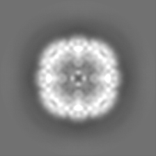
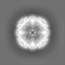
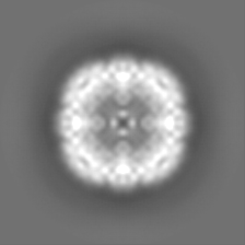
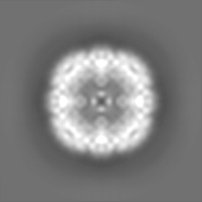
Movie
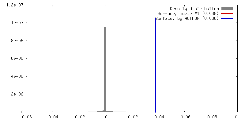
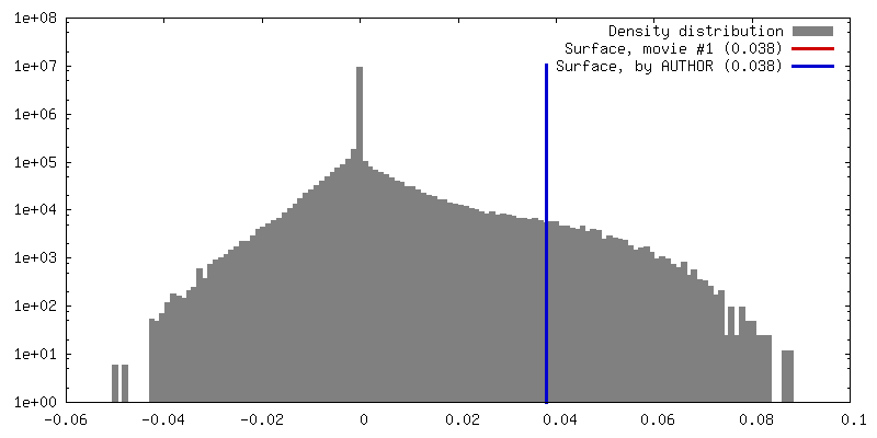
Surface view with section colored by density value
In the structure databanks used in Yorodumi, some data are registered as the other names, "COVID-19 virus" and "2019-nCoV". Here are the details of the virus and the list of structure data.
Jan 31, 2019. EMDB accession codes are about to change! (news from PDBe EMDB page)
EMDB accession codes are about to change! (news from PDBe EMDB page)
The allocation of 4 digits for EMDB accession codes will soon come to an end. Whilst these codes will remain in use, new EMDB accession codes will include an additional digit and will expand incrementally as the available range of codes is exhausted. The current 4-digit format prefixed with “EMD-” (i.e. EMD-XXXX) will advance to a 5-digit format (i.e. EMD-XXXXX), and so on. It is currently estimated that the 4-digit codes will be depleted around Spring 2019, at which point the 5-digit format will come into force.
The EM Navigator/Yorodumi systems omit the EMD- prefix.
Related info.:Q: What is EMD? / ID/Accession-code notation in Yorodumi/EM Navigator
Yorodumi is a browser for structure data from EMDB, PDB, SASBDB, etc.
This page is also the successor to EM Navigator detail page, and also detail information page/front-end page for Omokage search.
The word "yorodu" (or yorozu) is an old Japanese word meaning "ten thousand". "mi" (miru) is to see.
Related info.:EMDB / PDB / SASBDB / Comparison of 3 databanks / Yorodumi Search / Aug 31, 2016. New EM Navigator & Yorodumi / Yorodumi Papers / Jmol/JSmol / Function and homology information / Changes in new EM Navigator and Yorodumi
 Movie
Movie Controller


 Open data
 Basic information
 Map data
 Sample
 Function and homology information
 Authors
 United States, 1 items
  Citation
 Structure visualization
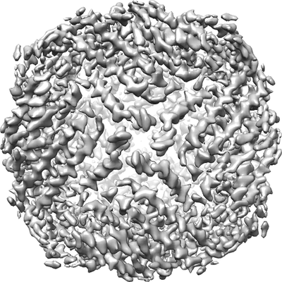
 Downloads & links
 emd_20157.png
 http://ftp.pdbj.org/pub/emdb/structures/EMD-20157


 Z (Sec.)
 Y (Row.)
 X (Col.)


 Sample components
 Processing
 Electron microscopy
 FIELD EMISSION GUN