 Movie
Movie Controller

[English] 日本語
 Yorodumi
- EMDB-10512: Cryo-EM reconstruction of TypeI tau filaments extracted from the ... -
+ Open data

- Basic information
| Entry | Database: EMDB / ID: EMD-10512 | |||||||||
|---|---|---|---|---|---|---|---|---|---|---|
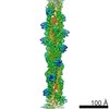
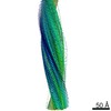
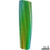
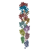
| Title | Cryo-EM reconstruction of TypeI tau filaments extracted from the brains of an individual (Case 1) with Corticobasal degeneration | |||||||||
 Map data Map data | Cryo-EM reconstruction of TypeI tau filaments extracted from the brains of an individual (Case 1) with Corticobasal degeneration | |||||||||
 Sample Sample |
| |||||||||
 Keywords Keywords | tau protein / filament / cross-beta structure / PROTEIN FIBRIL | |||||||||
| Function / homology |  Function and homology information Function and homology informationplus-end-directed organelle transport along microtubule / histone-dependent DNA binding / negative regulation of protein localization to mitochondrion / neurofibrillary tangle / microtubule lateral binding / axonal transport / tubulin complex / positive regulation of protein localization to synapse / phosphatidylinositol bisphosphate binding / generation of neurons ...plus-end-directed organelle transport along microtubule / histone-dependent DNA binding / negative regulation of protein localization to mitochondrion / neurofibrillary tangle / microtubule lateral binding / axonal transport / tubulin complex / positive regulation of protein localization to synapse / phosphatidylinositol bisphosphate binding / generation of neurons / rRNA metabolic process / axonal transport of mitochondrion / regulation of mitochondrial fission / axon development / regulation of microtubule-based movement / intracellular distribution of mitochondria / regulation of chromosome organization / central nervous system neuron development / minor groove of adenine-thymine-rich DNA binding / lipoprotein particle binding / microtubule polymerization / negative regulation of mitochondrial membrane potential / regulation of microtubule polymerization / dynactin binding / apolipoprotein binding / protein polymerization / main axon / Caspase-mediated cleavage of cytoskeletal proteins / regulation of microtubule polymerization or depolymerization / negative regulation of mitochondrial fission / axolemma / glial cell projection / neurofibrillary tangle assembly / positive regulation of axon extension / regulation of cellular response to heat / positive regulation of microtubule polymerization / positive regulation of protein localization / Activation of AMPK downstream of NMDARs / positive regulation of superoxide anion generation / cellular response to brain-derived neurotrophic factor stimulus / regulation of long-term synaptic depression / supramolecular fiber organization / cytoplasmic microtubule organization / regulation of calcium-mediated signaling / axon cytoplasm / somatodendritic compartment / synapse assembly / phosphatidylinositol binding / nuclear periphery / astrocyte activation / enzyme inhibitor activity / protein phosphatase 2A binding / stress granule assembly / regulation of microtubule cytoskeleton organization / regulation of autophagy / cellular response to reactive oxygen species / microglial cell activation / cellular response to nerve growth factor stimulus / Hsp90 protein binding / protein homooligomerization / SH3 domain binding / PKR-mediated signaling / synapse organization / regulation of synaptic plasticity / response to lead ion / microtubule cytoskeleton organization / memory / neuron projection development / cytoplasmic ribonucleoprotein granule / cell-cell signaling / single-stranded DNA binding / cellular response to heat / growth cone / protein-folding chaperone binding / microtubule cytoskeleton / actin binding / cell body / double-stranded DNA binding / sequence-specific DNA binding / microtubule binding / amyloid fibril formation / dendritic spine / microtubule / protein-macromolecule adaptor activity / learning or memory / neuron projection / membrane raft / negative regulation of gene expression / axon / neuronal cell body / DNA damage response / dendrite / protein kinase binding / enzyme binding / mitochondrion / DNA binding / RNA binding / extracellular region / identical protein binding / nucleus Similarity search - Function | |||||||||
| Biological species |  Homo sapiens (human) Homo sapiens (human) | |||||||||
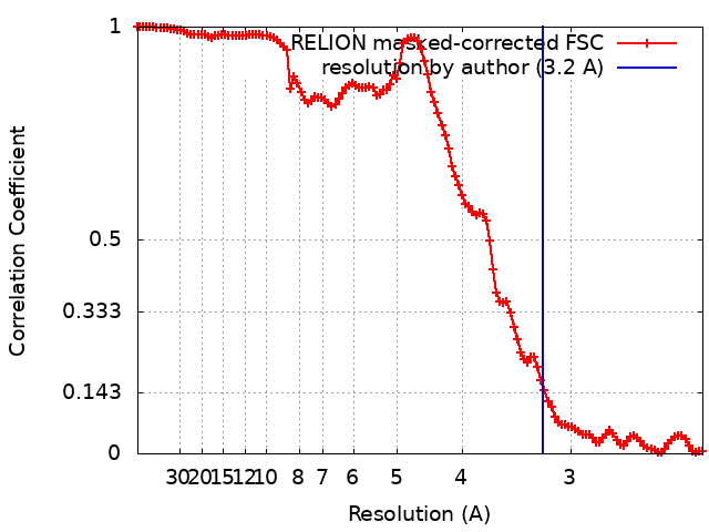
| Method | helical reconstruction / cryo EM / Resolution: 3.2 Å | |||||||||
 Authors Authors | Zhang W / Murzin AG / Falcon B / Shi Y / Goedert M / Scheres SHW | |||||||||
| Funding support |  United Kingdom, 2 items United Kingdom, 2 items
| |||||||||
 Citation Citation | Journal: Nature / Year: 2020 Title: Novel tau filament fold in corticobasal degeneration. Authors: Wenjuan Zhang / Airi Tarutani / Kathy L Newell / Alexey G Murzin / Tomoyasu Matsubara / Benjamin Falcon / Ruben Vidal / Holly J Garringer / Yang Shi / Takeshi Ikeuchi / Shigeo Murayama / ...Authors: Wenjuan Zhang / Airi Tarutani / Kathy L Newell / Alexey G Murzin / Tomoyasu Matsubara / Benjamin Falcon / Ruben Vidal / Holly J Garringer / Yang Shi / Takeshi Ikeuchi / Shigeo Murayama / Bernardino Ghetti / Masato Hasegawa / Michel Goedert / Sjors H W Scheres /   Abstract: Corticobasal degeneration (CBD) is a neurodegenerative tauopathy-a class of disorders in which the tau protein forms insoluble inclusions in the brain-that is characterized by motor and cognitive ...Corticobasal degeneration (CBD) is a neurodegenerative tauopathy-a class of disorders in which the tau protein forms insoluble inclusions in the brain-that is characterized by motor and cognitive disturbances. The H1 haplotype of MAPT (the tau gene) is present in cases of CBD at a higher frequency than in controls, and genome-wide association studies have identified additional risk factors. By histology, astrocytic plaques are diagnostic of CBD; by SDS-PAGE, so too are detergent-insoluble, 37 kDa fragments of tau. Like progressive supranuclear palsy, globular glial tauopathy and argyrophilic grain disease, CBD is characterized by abundant filamentous tau inclusions that are made of isoforms with four microtubule-binding repeats. This distinguishes such '4R' tauopathies from Pick's disease (the filaments of which are made of three-repeat (3R) tau isoforms) and from Alzheimer's disease and chronic traumatic encephalopathy (CTE) (in which both 3R and 4R isoforms are found in the filaments). Here we use cryo-electron microscopy to analyse the structures of tau filaments extracted from the brains of three individuals with CBD. These filaments were identical between cases, but distinct from those seen in Alzheimer's disease, Pick's disease and CTE. The core of a CBD filament comprises residues lysine 274 to glutamate 380 of tau, spanning the last residue of the R1 repeat, the whole of the R2, R3 and R4 repeats, and 12 amino acids after R4. The core adopts a previously unseen four-layered fold, which encloses a large nonproteinaceous density. This density is surrounded by the side chains of lysine residues 290 and 294 from R2 and lysine 370 from the sequence after R4. | |||||||||
| History |
|
- Structure visualization
| Movie |
Movie viewer |
|---|---|
| Structure viewer | EM map: SurfViewMolmilJmol/JSmol |
| Supplemental images |

- Downloads & links
-EMDB archive
| Map data | emd_10512.map.gz | 16.5 MB | EMDB map data format | |
|---|---|---|---|---|
| Header (meta data) | emd-10512-v30.xmlemd-10512.xml | 20.5 KB 20.5 KB | Display Display | EMDB header |
| FSC (resolution estimation) | emd_10512_fsc.xml | 11.7 KB | Display | FSC data file |
| Images | 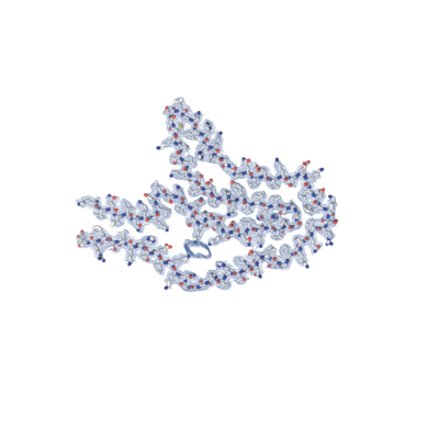 emd_10512.png emd_10512.png | 59.5 KB | ||
| Masks | emd_10512_msk_1.map | 137.1 MB | Mask map | |
| Filedesc metadata | emd-10512.cif.gz | 7 KB | ||
| Others | emd_10512_half_map_1.map.gzemd_10512_half_map_2.map.gz | 16.4 MB 16.4 MB | ||
| Archive directory |  http://ftp.pdbj.org/pub/emdb/structures/EMD-10512ftp://ftp.pdbj.org/pub/emdb/structures/EMD-10512 http://ftp.pdbj.org/pub/emdb/structures/EMD-10512ftp://ftp.pdbj.org/pub/emdb/structures/EMD-10512 | HTTPS FTP |
-Related structure data
| Related structure data |  6tjoMC  6tjxC C: citing same article ( M: atomic model generated by this map |
|---|---|
| Similar structure data | |
| EM raw data | EMPIAR-10340 (Title: Cryo-EM reconstruction of tau filaments extracted from the brains of three individuals with Corticobasal degeneration Data size: 2.8 TB Data #1: Unaligned movies for Case 1 [micrographs - multiframe] Data #2: Unaligned movies for Case 2 [micrographs - multiframe] Data #3: Unaligned movies for Case 3 [micrographs - multiframe]) |

-Links
| EMDB pages | EMDB (EBI/PDBe) / EMDataResource |
|---|---|
| Related items in Molecule of the Month |


-Map
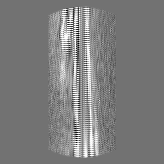
| File | Download / File: emd_10512.map.gz / Format: CCP4 / Size: 137.1 MB / Type: IMAGE STORED AS FLOATING POINT NUMBER (4 BYTES) | ||||||||||||||||||||||||||||||||||||||||||||||||||||||||||||
|---|---|---|---|---|---|---|---|---|---|---|---|---|---|---|---|---|---|---|---|---|---|---|---|---|---|---|---|---|---|---|---|---|---|---|---|---|---|---|---|---|---|---|---|---|---|---|---|---|---|---|---|---|---|---|---|---|---|---|---|---|---|
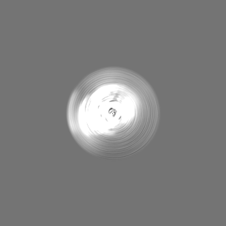
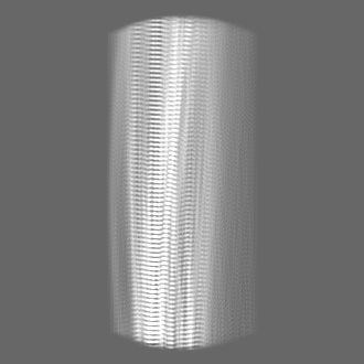
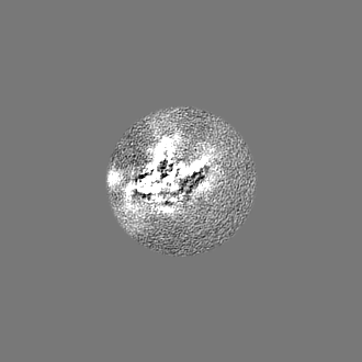
| Annotation | Cryo-EM reconstruction of TypeI tau filaments extracted from the brains of an individual (Case 1) with Corticobasal degeneration | ||||||||||||||||||||||||||||||||||||||||||||||||||||||||||||
| Projections & slices | Image control
Images are generated by Spider. | ||||||||||||||||||||||||||||||||||||||||||||||||||||||||||||
| Voxel size | X=Y=Z: 1.15 Å | ||||||||||||||||||||||||||||||||||||||||||||||||||||||||||||
| Density |
| ||||||||||||||||||||||||||||||||||||||||||||||||||||||||||||
| Symmetry | Space group: 1 | ||||||||||||||||||||||||||||||||||||||||||||||||||||||||||||
| Details | EMDB XML:
CCP4 map header:
| ||||||||||||||||||||||||||||||||||||||||||||||||||||||||||||
 Z (Sec.)
 Y (Row.)
 X (Col.)


-Supplemental data
-Mask #1
| File | emd_10512_msk_1.map | ||||||||||||
|---|---|---|---|---|---|---|---|---|---|---|---|---|---|
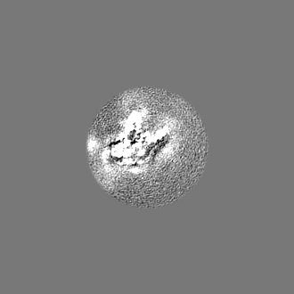
| Projections & Slices |
| ||||||||||||
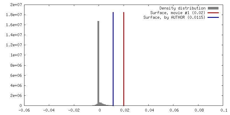
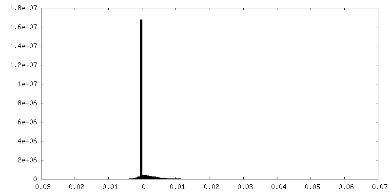
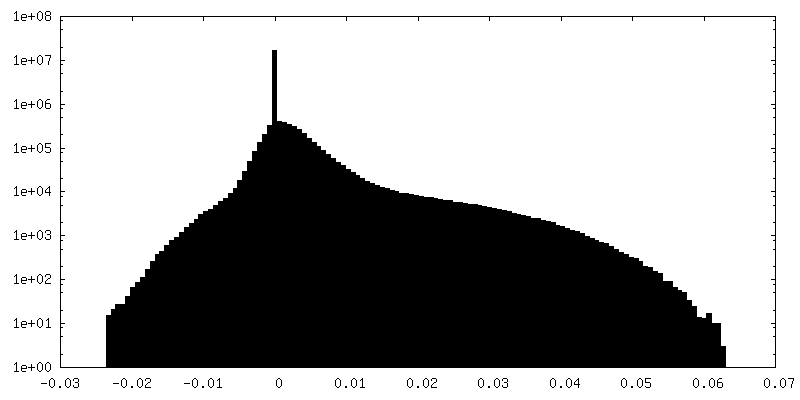
| Density Histograms |


-Half map: Half map 1
| File | emd_10512_half_map_1.map | ||||||||||||
|---|---|---|---|---|---|---|---|---|---|---|---|---|---|
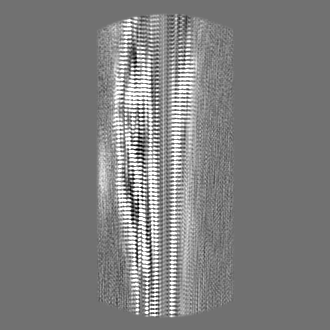
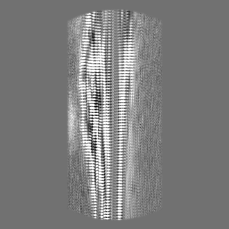
| Annotation | Half map 1 | ||||||||||||
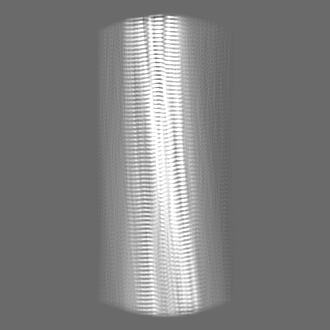
| Projections & Slices |
| ||||||||||||
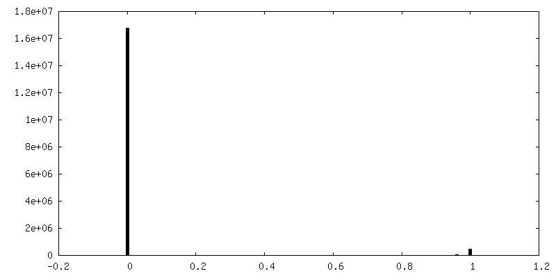
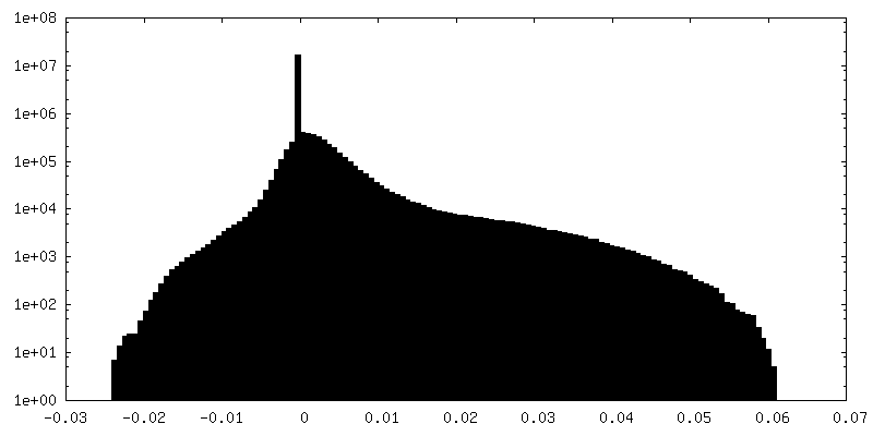
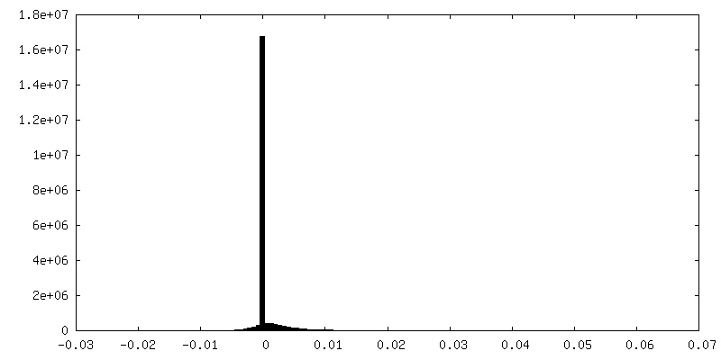
| Density Histograms |

-Half map: Half map 2
| File | emd_10512_half_map_2.map | ||||||||||||
|---|---|---|---|---|---|---|---|---|---|---|---|---|---|
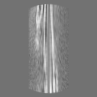
| Annotation | Half map 2 | ||||||||||||
| Projections & Slices |
| ||||||||||||
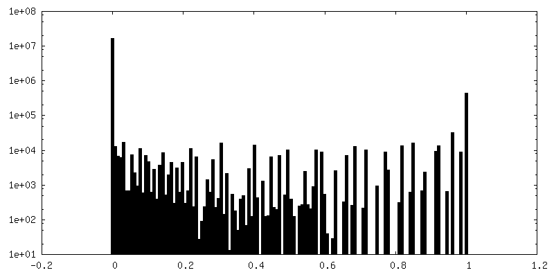
| Density Histograms |


- Sample components
-Entire : Tau filaments extracted from the Frontal cortex of a patient with...
| Entire | Name: Tau filaments extracted from the Frontal cortex of a patient with corticobasal degeneration. |
|---|---|
| Components |
|
-Supramolecule #1: Tau filaments extracted from the Frontal cortex of a patient with...
| Supramolecule | Name: Tau filaments extracted from the Frontal cortex of a patient with corticobasal degeneration. type: complex / ID: 1 / Parent: 0 / Macromolecule list: all Details: Corticobasal degeneration (CBD) is characterised by abundant filamentous tau inclusions that are made of isoforms with four microtubule-binding repeats (4R). |
|---|---|
| Source (natural) | Organism: Homo sapiens (human) |
| Molecular weight | Theoretical: 460 KDa |
-Macromolecule #1: Microtubule-associated protein tau
| Macromolecule | Name: Microtubule-associated protein tau / type: protein_or_peptide / ID: 1 / Number of copies: 3 / Enantiomer: LEVO |
|---|---|
| Source (natural) | Organism: Homo sapiens (human) |
| Molecular weight | Theoretical: 45.919871 KDa |
| Sequence | String: MAEPRQEFEV MEDHAGTYGL GDRKDQGGYT MHQDQEGDTD AGLKESPLQT PTEDGSEEPG SETSDAKSTP TAEDVTAPLV DEGAPGKQA AAQPHTEIPE GTTAEEAGIG DTPSLEDEAA GHVTQARMVS KSKDGTGSDD KKAKGADGKT KIATPRGAAP P GQKGQANA ...String: MAEPRQEFEV MEDHAGTYGL GDRKDQGGYT MHQDQEGDTD AGLKESPLQT PTEDGSEEPG SETSDAKSTP TAEDVTAPLV DEGAPGKQA AAQPHTEIPE GTTAEEAGIG DTPSLEDEAA GHVTQARMVS KSKDGTGSDD KKAKGADGKT KIATPRGAAP P GQKGQANA TRIPAKTPPA PKTPPSSGEP PKSGDRSGYS SPGSPGTPGS RSRTPSLPTP PTREPKKVAV VRTPPKSPSS AK SRLQTAP VPMPDLKNVK SKIGSTENLK HQPGGGKVQI INKKLDLSNV QSKCGSKDNI KHVPGGGSVQ IVYKPVDLSK VTS KCGSLG NIHHKPGGGQ VEVKSEKLDF KDRVQSKIGS LDNITHVPGG GNKKIETHKL TFRENAKAKT DHGAEIVYKS PVVS GDTSP RHLSNVSSTG SIDMVDSPQL ATLADEVSAS LAKQGL UniProtKB: Microtubule-associated protein tau |
-Experimental details
-Structure determination
| Method | cryo EM |
|---|---|
 Processing Processing | helical reconstruction |
| Aggregation state | filament |
-Sample preparation
| Concentration | 2.0 mg/mL | |||||||||
|---|---|---|---|---|---|---|---|---|---|---|
| Buffer | pH: 7.4 Component:
Details: 20 mM Tris, pH 7.4, 100mM NaCl | |||||||||
| Vitrification | Cryogen name: ETHANE / Chamber humidity: 100 % / Chamber temperature: 277 K / Instrument: FEI VITROBOT MARK IV / Details: Blot force: -12 ; Blot time: 4s. | |||||||||
| Details | The tau filaments were extracted from the Frontal cortex of a patient with corticobasal degeneration by using sarkosyl and ultracentrifuge. |
- Electron microscopy
| Microscope | FEI TITAN KRIOS |
|---|---|
| Specialist optics | Energy filter - Name: GIF Quantum LS / Energy filter - Slit width: 20 eV |
| Image recording | Film or detector model: GATAN K2 SUMMIT (4k x 4k) / Detector mode: COUNTING / Digitization - Dimensions - Width: 3838 pixel / Digitization - Dimensions - Height: 3710 pixel / Digitization - Frames/image: 1-40 / Number grids imaged: 1 / Number real images: 2567 / Average exposure time: 10.0 sec. / Average electron dose: 1.346 e/Å2 Details: Images were collected in movie-mode at 40 frames every 10 seconds |
| Electron beam | Acceleration voltage: 300 kV / Electron source:  FIELD EMISSION GUN FIELD EMISSION GUN |
| Electron optics | C2 aperture diameter: 70.0 µm / Illumination mode: FLOOD BEAM / Imaging mode: BRIGHT FIELD / Cs: 2.7 mm / Nominal defocus max: 2.8000000000000003 µm / Nominal defocus min: 1.7 µm |
| Sample stage | Specimen holder model: FEI TITAN KRIOS AUTOGRID HOLDER / Cooling holder cryogen: NITROGEN |
| Experimental equipment |  Model: Titan Krios / Image courtesy: FEI Company |
+Image processing
-Atomic model buiding 1
| Details | A stack of three consecutive monomers was refined to preserve nearest-neighbour interactions for the middle chain. Because most residues adopted cross strand conformation, hydrogen-bond restraints were imposed to preserve a parallel, in-register hydrogen bonding pattern in earlier stages of Fourier-space refinements. Local symmetry restraints were imposed to keep all beta strand rungs identical. Side-chain clashes were detected using MOLPROBITY, and corrected by iterative cycles of real-space refinement in COOT and Fourier-space refinement in REFMAC and PHENIX. For each refined structure, separate model refinements were performed against a single half-map, and the resulting model was compared to the other half-map to confirm the absence of overfitting. |
|---|---|
| Refinement | Space: RECIPROCAL / Protocol: AB INITIO MODEL / Overall B value: 26.63 / Target criteria: Fourier shell correlation |
| Output model | PDB-6tjo: |
