 Movie
Movie Controller

[English] 日本語
 Yorodumi
- PDB-4aso: TubR bound to 24 bp of tubC from Bacillus thuringiensis serovar i... -
+ Open data

- Basic information
| Entry | Database: PDB / ID: 4aso | ||||||
|---|---|---|---|---|---|---|---|
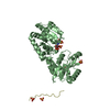
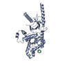
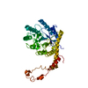
| Title | TubR bound to 24 bp of tubC from Bacillus thuringiensis serovar israelensis pBtoxis | ||||||
 Components Components |
| ||||||
 Keywords Keywords | STRUCTURAL PROTEIN/DNA / STRUCTURAL PROTEIN-DNA COMPLEX / PARTITION / SEGREGATION | ||||||
| Function / homology | plasmid partitioning / Winged helix DNA-binding domain superfamily / Winged helix-like DNA-binding domain superfamily / DNA binding / identical protein binding / DNA / DNA (> 10) / DNA-binding transcriptional repressor TubR Function and homology information Function and homology information | ||||||
| Biological species |  | ||||||
| Method |  X-RAY DIFFRACTION / SYNCHROTRON / MOLECULAR REPLACEMENT / Resolution: 7 Å X-RAY DIFFRACTION / SYNCHROTRON / MOLECULAR REPLACEMENT / Resolution: 7 Å | ||||||
 Authors Authors | Aylett, C.H.S. / Lowe, J. | ||||||
 Citation Citation | Journal: Proc.Natl.Acad.Sci.USA / Year: 2012 Title: Superstructure of the Centromeric Complex of Tubzrc Plasmid Partitioning Systems. Authors: Aylett, C.H.S. / Lowe, J. | ||||||
| History |
|
- Structure visualization
| Structure viewer | Molecule: MolmilJmol/JSmol |
|---|
- Downloads & links
-Download
| PDBx/mmCIF format | 4aso.cif.gz | 384.7 KB | Display | PDBx/mmCIF format |
|---|---|---|---|---|
| PDB format | pdb4aso.ent.gz | 307.8 KB | Display | PDB format |
| PDBx/mmJSON format | 4aso.json.gz | Tree view | PDBx/mmJSON format | |
| Others |  Other downloads Other downloads |
-Validation report
| Arichive directory | https://data.pdbj.org/pub/pdb/validation_reports/as/4asoftp://data.pdbj.org/pub/pdb/validation_reports/as/4aso | HTTPS FTP |
|---|
-Related structure data
| Related structure data |  4asnC  4assC  3m9aS C: citing same article ( S: Starting model for refinement |
|---|---|
| Similar structure data |


-Links
 PDBj
PDBj


- Assembly
| Deposited unit | 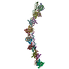
| ||||||||
|---|---|---|---|---|---|---|---|---|---|
| 1 | 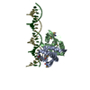
| ||||||||
| 2 | 
| ||||||||
| 3 | 
| ||||||||
| 4 | 
| ||||||||
| 5 | 
| ||||||||
| 6 | 
| ||||||||
| 7 | 
| ||||||||
| 8 | 
| ||||||||
| Unit cell |
|
-Components
| #1: Protein | Mass: 12119.912 Da / Num. of mol.: 16 Source method: isolated from a genetically manipulated source Source: (gene. exp.) #2: DNA chain | Mass: 7323.761 Da / Num. of mol.: 4 / Fragment: SENSE STRAND / Source method: obtained synthetically / Source: (synth.) #3: DNA chain | Mass: 7408.855 Da / Num. of mol.: 4 / Fragment: ANTISENSE STRAND / Source method: obtained synthetically / Source: (synth.) Has protein modification | Y | Sequence details | BP 126549-126514 DNA IS CONTINUOUS IN THE CRYSTALS, BUT HAS BEEN CUT FOR REFINEMENT TO CENTRE THE ...BP 126549-126514 DNA IS CONTINUOUS | |
|---|
-Experimental details
-Experiment
| Experiment | Method: X-RAY DIFFRACTION / Number of used crystals: 1 |
|---|
- Sample preparation
| Crystal | Density Matthews: 4.81 Å3/Da / Density % sol: 74.54 % Description: RIGID BODY ON VERY LOW RESOLUTION DATA. STRUCTURE CONFIRMED BY MSE SAD ANOMALOUS DIFFERENCE MAP. REGISTER OF DNA WAS INFERRED BY MICROARRAY EXPERIMENT. |
|---|---|
| Crystal grow | pH: 7.5 Details: BT TUBR-TUBC CRYSTALS WERE PRODUCED IN 500 NL TO 500 NL PROTEIN TO PRECIPITANT DROPS: 10 MG/ML BT TUBR, 500 UM TUBC-24, 100 MM NA-HEPES PH 7.5, 5 MM MGCL2, 25 % (W/V) POLYACRYLIC ACID 5100 SODIUM SALT. |
-Data collection
| Diffraction | Mean temperature: 100 K |
|---|---|
| Diffraction source | Source: SYNCHROTRON / Site: Diamond  / Beamline: I03 / Wavelength: 0.9794 / Beamline: I03 / Wavelength: 0.9794 |
| Detector | Type: DECTRIS PILATUS 6M / Detector: PIXEL / Date: May 29, 2011 |
| Radiation | Monochromator: GRAPHITE CRYSTAL / Protocol: SINGLE WAVELENGTH / Monochromatic (M) / Laue (L): M / Scattering type: x-ray |
| Radiation wavelength | Wavelength: 0.9794 Å / Relative weight: 1 |
| Reflection | Resolution: 7→257.87 Å / Num. obs: 8779 / % possible obs: 98.1 % / Observed criterion σ(I): 1.3 / Redundancy: 5.9 % / Rmerge(I) obs: 0.09 / Net I/σ(I): 10.9 |
| Reflection shell | Resolution: 7→7.38 Å / Redundancy: 5.9 % / Rmerge(I) obs: 1.642 / Mean I/σ(I) obs: 1.3 / % possible all: 98.5 |
- Processing
| Software |
| |||||||||||||||||||||||||||||||||||||||||||||||||
|---|---|---|---|---|---|---|---|---|---|---|---|---|---|---|---|---|---|---|---|---|---|---|---|---|---|---|---|---|---|---|---|---|---|---|---|---|---|---|---|---|---|---|---|---|---|---|---|---|---|---|
| Refinement | Method to determine structure: MOLECULAR REPLACEMENT Starting model: PDB ENTRY 3M9A Resolution: 7→64.469 Å / SU ML: 1.48 / σ(F): 1.34 / Phase error: 42.71 / Stereochemistry target values: ML
| |||||||||||||||||||||||||||||||||||||||||||||||||
| Solvent computation | Shrinkage radii: 0.9 Å / VDW probe radii: 1.11 Å / Solvent model: FLAT BULK SOLVENT MODEL | |||||||||||||||||||||||||||||||||||||||||||||||||
| Refinement step | Cycle: LAST / Resolution: 7→64.469 Å
| |||||||||||||||||||||||||||||||||||||||||||||||||
| Refine LS restraints |
| |||||||||||||||||||||||||||||||||||||||||||||||||
| LS refinement shell |
|
