 Movie
Movie Controller

[English] 日本語
 Yorodumi
- EMDB-10362: Cryo-EM structure of the wild-type flagellar filament of the Firm... -
+ Open data

- Basic information
| Entry | Database: EMDB / ID: EMD-10362 | |||||||||||||||
|---|---|---|---|---|---|---|---|---|---|---|---|---|---|---|---|---|
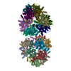
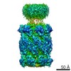
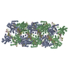
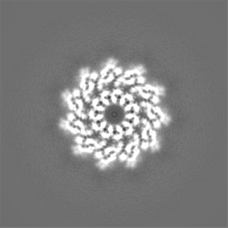
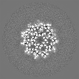
| Title | Cryo-EM structure of the wild-type flagellar filament of the Firmicute Kurthia | |||||||||||||||
 Map data Map data | ||||||||||||||||
 Sample Sample |
| |||||||||||||||
 Keywords Keywords | Gram-Positive Bacteria Flagella / Helical / Wild-Type / protein fibril | |||||||||||||||
| Function / homology |  Function and homology information Function and homology informationbacterial-type flagellum / structural molecule activity / extracellular region Similarity search - Function | |||||||||||||||
| Biological species |  Kurthia sp. 11kri321 (bacteria) Kurthia sp. 11kri321 (bacteria) | |||||||||||||||
| Method | helical reconstruction / cryo EM / Resolution: 2.8 Å | |||||||||||||||
 Authors Authors | Blum TB / Abrahams JP | |||||||||||||||
| Funding support |  Switzerland, 4 items Switzerland, 4 items
| |||||||||||||||
 Citation Citation | Journal: Sci Rep / Year: 2019 Title: The wild-type flagellar filament of the Firmicute Kurthia at 2.8 Å resolution in vivo. Authors: Thorsten B Blum / Sevasti Filippidou / Mathilda Fatton / Pilar Junier / Jan Pieter Abrahams /  Abstract: Bacteria swim and swarm by rotating the micrometers long, helical filaments of their flagella. They change direction by reversing their flagellar rotation, which switches the handedness of the ...Bacteria swim and swarm by rotating the micrometers long, helical filaments of their flagella. They change direction by reversing their flagellar rotation, which switches the handedness of the filament's supercoil. So far, all studied functional filaments are composed of a mixture of L- and R-state flagellin monomers. Here we show in a study of the wild type Firmicute Kurthia sp., that curved, functional filaments can adopt a conformation in vivo that is closely related to a uniform, all-L-state. This sheds additional light on transitions of the flagellar supercoil and uniquely reveals the atomic structure of a wild-type flagellar filament in vivo, including six residues showing clearly densities of O-linked glycosylation. | |||||||||||||||
| History |
|
- Structure visualization
| Movie |
Movie viewer |
|---|---|
| Structure viewer | EM map: SurfViewMolmilJmol/JSmol |
| Supplemental images |

- Downloads & links
-EMDB archive
| Map data | emd_10362.map.gz | 57.4 MB | EMDB map data format | |
|---|---|---|---|---|
| Header (meta data) | emd-10362-v30.xmlemd-10362.xml | 9.9 KB 9.9 KB | Display Display | EMDB header |
| FSC (resolution estimation) | emd_10362_fsc.xml | 9.1 KB | Display | FSC data file |
| Images | 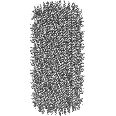 emd_10362.png emd_10362.png | 65.8 KB | ||
| Filedesc metadata | emd-10362.cif.gz | 4.9 KB | ||
| Archive directory |  http://ftp.pdbj.org/pub/emdb/structures/EMD-10362ftp://ftp.pdbj.org/pub/emdb/structures/EMD-10362 http://ftp.pdbj.org/pub/emdb/structures/EMD-10362ftp://ftp.pdbj.org/pub/emdb/structures/EMD-10362 | HTTPS FTP |
-Related structure data
| Related structure data |  6t17MC M: atomic model generated by this map C: citing same article ( |
|---|---|
| Similar structure data |

-Links
| EMDB pages | EMDB (EBI/PDBe) / EMDataResource |
|---|
-Map
| File | Download / File: emd_10362.map.gz / Format: CCP4 / Size: 64 MB / Type: IMAGE STORED AS FLOATING POINT NUMBER (4 BYTES) | ||||||||||||||||||||||||||||||||||||||||||||||||||||||||||||||||||||
|---|---|---|---|---|---|---|---|---|---|---|---|---|---|---|---|---|---|---|---|---|---|---|---|---|---|---|---|---|---|---|---|---|---|---|---|---|---|---|---|---|---|---|---|---|---|---|---|---|---|---|---|---|---|---|---|---|---|---|---|---|---|---|---|---|---|---|---|---|---|
| Projections & slices | Image control
Images are generated by Spider. | ||||||||||||||||||||||||||||||||||||||||||||||||||||||||||||||||||||
| Voxel size | X=Y=Z: 1.058 Å | ||||||||||||||||||||||||||||||||||||||||||||||||||||||||||||||||||||
| Density |
| ||||||||||||||||||||||||||||||||||||||||||||||||||||||||||||||||||||
| Symmetry | Space group: 1 | ||||||||||||||||||||||||||||||||||||||||||||||||||||||||||||||||||||
| Details | EMDB XML:
CCP4 map header:
| ||||||||||||||||||||||||||||||||||||||||||||||||||||||||||||||||||||
 Z (Sec.)
 Y (Row.)
 X (Col.)


-Supplemental data
- Sample components
-Entire : Flagellar Filament
| Entire | Name: Flagellar Filament |
|---|---|
| Components |
|
-Supramolecule #1: Flagellar Filament
| Supramolecule | Name: Flagellar Filament / type: complex / ID: 1 / Parent: 0 / Macromolecule list: all |
|---|---|
| Source (natural) | Organism: Kurthia sp. 11kri321 (bacteria) |
-Macromolecule #1: Flagellin
| Macromolecule | Name: Flagellin / type: protein_or_peptide / ID: 1 / Number of copies: 44 / Enantiomer: LEVO |
|---|---|
| Source (natural) | Organism: Kurthia sp. 11kri321 (bacteria) |
| Molecular weight | Theoretical: 29.530055 KDa |
| Recombinant expression | Organism: Kurthia sp. 11kri321 (bacteria) |
| Sequence | String: MRIQHNIAAL NTHRNLAANN AAASKNLEKL SSGFKINRAG DDAAGLAISE KMRGQISGLN MASKNSSDAI SLIQTAEGGL NETHAILQR MRELAVQSRN DTNDEATNDR SNLNDELKQL QEEITRISSQ MEFNNKKLLD GSQSTNGLTF QIGANAGQTI T MKISTMSA ...String: MRIQHNIAAL NTHRNLAANN AAASKNLEKL SSGFKINRAG DDAAGLAISE KMRGQISGLN MASKNSSDAI SLIQTAEGGL NETHAILQR MRELAVQSRN DTNDEATNDR SNLNDELKQL QEEITRISSQ MEFNNKKLLD GSQSTNGLTF QIGANAGQTI T MKISTMSA TKLGVDAAKA SISKGTAASK AIKSIDDAIN TVSKTRSALG AVQNRLEHTI NNLGTSAENL TAAESRIRDT DM AAEMMAF TKNNILTQAA QSMLAQANQQ PQGVLQLLQ UniProtKB: Flagellin |
-Experimental details
-Structure determination
| Method | cryo EM |
|---|---|
 Processing Processing | helical reconstruction |
| Aggregation state | filament |
-Sample preparation
| Buffer | pH: 7.3 |
|---|---|
| Vitrification | Cryogen name: ETHANE / Instrument: LEICA EM GP |
- Electron microscopy
| Microscope | FEI TITAN KRIOS |
|---|---|
| Image recording | Film or detector model: GATAN K2 SUMMIT (4k x 4k) / Detector mode: SUPER-RESOLUTION / Average electron dose: 86.0 e/Å2 |
| Electron beam | Acceleration voltage: 300 kV / Electron source:  FIELD EMISSION GUN FIELD EMISSION GUN |
| Electron optics | Illumination mode: FLOOD BEAM / Imaging mode: BRIGHT FIELD |
| Experimental equipment |  Model: Titan Krios / Image courtesy: FEI Company |
-Image processing
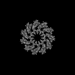
| Final reconstruction | Number classes used: 1 Applied symmetry - Helical parameters - Δz: 4.83 Å Applied symmetry - Helical parameters - Δ&Phi: 65.45 ° Applied symmetry - Helical parameters - Axial symmetry: C1 (asymmetric) Algorithm: BACK PROJECTION / Resolution.type: BY AUTHOR / Resolution: 2.8 Å / Resolution method: FSC 0.143 CUT-OFF / Number images used: 9270 |
|---|---|
| Startup model | Type of model: OTHER / Details: Sub-tomogram average |
| Final angle assignment | Type: NOT APPLICABLE |
| FSC plot (resolution estimation) | 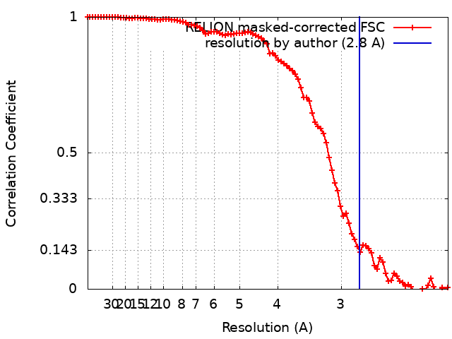 |
