Movie
Movie Controller


+ Open data

- Basic information
| Entry | Database: EMDB / ID: EMD-10373 | |||||||||
|---|---|---|---|---|---|---|---|---|---|---|
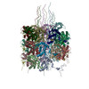
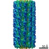
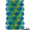
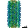
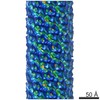
| Title | CryoEM structure for Turnip mosaic virus (TuMV) | |||||||||
 Map data Map data | CryoEM map for Turnip mosaic virus (TuMV) | |||||||||
 Sample Sample |
| |||||||||
 Keywords Keywords | potyvirus / virion / helical virus / VIRUS | |||||||||
| Function / homology | Potyvirus coat protein / Potyvirus coat protein / viral capsid / Genome polyprotein / Genome polyprotein Function and homology information Function and homology information | |||||||||
| Biological species |  Turnip mosaic virus (strain Japanese) / Turnip mosaic virus Turnip mosaic virus (strain Japanese) / Turnip mosaic virus | |||||||||
| Method | helical reconstruction / cryo EM / Resolution: 5.2 Å | |||||||||
 Authors Authors | Valle MV / Cuesta R | |||||||||
| Funding support |  Spain, 2 items Spain, 2 items
| |||||||||
 Citation Citation | Journal: Sci Rep / Year: 2019 Title: Structure of Turnip mosaic virus and its viral-like particles. Authors: Rebeca Cuesta / Carmen Yuste-Calvo / David Gil-Cartón / Flora Sánchez / Fernando Ponz / Mikel Valle / Abstract: Turnip mosaic virus (TuMV), a potyvirus, is a flexible filamentous plant virus that displays a helical arrangement of coat protein copies (CPs) bound to the ssRNA genome. TuMV is a bona fide ...Turnip mosaic virus (TuMV), a potyvirus, is a flexible filamentous plant virus that displays a helical arrangement of coat protein copies (CPs) bound to the ssRNA genome. TuMV is a bona fide representative of the Potyvirus genus, one of most abundant groups of plant viruses, which displays a very wide host range. We have studied by cryoEM the structure of TuMV virions and its viral-like particles (VLPs) to explore the role of the interactions between proteins and RNA in the assembly of the virions. The results show that the CP-RNA interaction is needed for the correct orientation of the CP N-terminal arm, a region that plays as a molecular staple between CP subunits in the fully assembled virion. | |||||||||
| History |
|
- Structure visualization
| Movie |
Movie viewer |
|---|---|
| Structure viewer | EM map: SurfViewMolmilJmol/JSmol |
| Supplemental images |

- Downloads & links
-EMDB archive
| Map data | emd_10373.map.gz | 19.6 MB | EMDB map data format | |
|---|---|---|---|---|
| Header (meta data) | emd-10373-v30.xmlemd-10373.xml | 12.3 KB 12.3 KB | Display Display | EMDB header |
| Images | 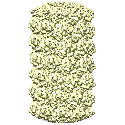 emd_10373.png emd_10373.png | 197.4 KB | ||
| Filedesc metadata | emd-10373.cif.gz | 5.5 KB | ||
| Archive directory |  http://ftp.pdbj.org/pub/emdb/structures/EMD-10373ftp://ftp.pdbj.org/pub/emdb/structures/EMD-10373 http://ftp.pdbj.org/pub/emdb/structures/EMD-10373ftp://ftp.pdbj.org/pub/emdb/structures/EMD-10373 | HTTPS FTP |
-Related structure data
| Related structure data |  6t34MC M: atomic model generated by this map C: citing same article ( |
|---|---|
| Similar structure data |


-Links
| EMDB pages | EMDB (EBI/PDBe) / EMDataResource |
|---|
-Map
| File | Download / File: emd_10373.map.gz / Format: CCP4 / Size: 59.6 MB / Type: IMAGE STORED AS FLOATING POINT NUMBER (4 BYTES) | ||||||||||||||||||||||||||||||||||||||||||||||||||||||||||||||||||||
|---|---|---|---|---|---|---|---|---|---|---|---|---|---|---|---|---|---|---|---|---|---|---|---|---|---|---|---|---|---|---|---|---|---|---|---|---|---|---|---|---|---|---|---|---|---|---|---|---|---|---|---|---|---|---|---|---|---|---|---|---|---|---|---|---|---|---|---|---|---|
| Annotation | CryoEM map for Turnip mosaic virus (TuMV) | ||||||||||||||||||||||||||||||||||||||||||||||||||||||||||||||||||||
| Projections & slices | Image control
Images are generated by Spider. | ||||||||||||||||||||||||||||||||||||||||||||||||||||||||||||||||||||
| Voxel size | X=Y=Z: 1.1 Å | ||||||||||||||||||||||||||||||||||||||||||||||||||||||||||||||||||||
| Density |
| ||||||||||||||||||||||||||||||||||||||||||||||||||||||||||||||||||||
| Symmetry | Space group: 1 | ||||||||||||||||||||||||||||||||||||||||||||||||||||||||||||||||||||
| Details | EMDB XML:
CCP4 map header:
| ||||||||||||||||||||||||||||||||||||||||||||||||||||||||||||||||||||
 Z (Sec.)
 Y (Row.)
 X (Col.)


-Supplemental data
- Sample components
-Entire : Turnip mosaic virus
| Entire | Name: Turnip mosaic virus |
|---|---|
| Components |
|
-Supramolecule #1: Turnip mosaic virus
| Supramolecule | Name: Turnip mosaic virus / type: virus / ID: 1 / Parent: 0 / Macromolecule list: all / NCBI-ID: 12230 / Sci species name: Turnip mosaic virus / Virus type: VIRION / Virus isolate: SPECIES / Virus enveloped: No / Virus empty: No |
|---|---|
| Host (natural) | Organism:  |
-Macromolecule #1: Coat protein
| Macromolecule | Name: Coat protein / type: protein_or_peptide / ID: 1 / Number of copies: 19 / Enantiomer: LEVO |
|---|---|
| Source (natural) | Organism: Turnip mosaic virus (strain Japanese) / Strain: Japanese |
| Molecular weight | Theoretical: 24.073395 KDa |
| Sequence | String: PRLKSLTSKM RVPRYEKRVA LNLDHLILYT PEQTDLSNTR STRKQFDTWF EGVMADYELT EDKMQIILNG LMVWCIENGT SPNINGMWV MMDGDDQVEF PIKPLIDHAK PTFRQIMAHF SDVAEAYIEK RNQDRPYMPR YGLQRNLTDM SLARYAFDFY E MTSRTPIR ...String: PRLKSLTSKM RVPRYEKRVA LNLDHLILYT PEQTDLSNTR STRKQFDTWF EGVMADYELT EDKMQIILNG LMVWCIENGT SPNINGMWV MMDGDDQVEF PIKPLIDHAK PTFRQIMAHF SDVAEAYIEK RNQDRPYMPR YGLQRNLTDM SLARYAFDFY E MTSRTPIR AREAHIQMKA AALRGANNNL FGLDGNVGTT VENTERHTT UniProtKB: Genome polyprotein |
-Macromolecule #2: RNA (5'-R(P*UP*UP*UP*UP*U)-3')
| Macromolecule | Name: RNA (5'-R(P*UP*UP*UP*UP*U)-3') / type: rna / ID: 2 / Details: Model of ssRNA as polyU / Number of copies: 19 |
|---|---|
| Source (natural) | Organism: Turnip mosaic virus |
| Molecular weight | Theoretical: 1.485872 KDa |
| Sequence | String: UUUUU |
-Experimental details
-Structure determination
| Method | cryo EM |
|---|---|
 Processing Processing | helical reconstruction |
| Aggregation state | filament |
-Sample preparation
| Buffer | pH: 7.5 |
|---|---|
| Vitrification | Cryogen name: ETHANE |
- Electron microscopy
| Microscope | FEI TITAN KRIOS |
|---|---|
| Image recording | Film or detector model: GATAN K2 SUMMIT (4k x 4k) / Detector mode: COUNTING / Digitization - Frames/image: 3-31 / Number grids imaged: 1 / Average exposure time: 9.0 sec. / Average electron dose: 40.0 e/Å2 |
| Electron beam | Acceleration voltage: 300 kV / Electron source:  FIELD EMISSION GUN FIELD EMISSION GUN |
| Electron optics | Illumination mode: FLOOD BEAM / Imaging mode: BRIGHT FIELD |
| Sample stage | Specimen holder model: FEI TITAN KRIOS AUTOGRID HOLDER / Cooling holder cryogen: NITROGEN |
| Experimental equipment |  Model: Titan Krios / Image courtesy: FEI Company |
-Image processing
| Final reconstruction | Applied symmetry - Helical parameters - Δz: 4.0 Å Applied symmetry - Helical parameters - Δ&Phi: -40.8 ° Applied symmetry - Helical parameters - Axial symmetry: C1 (asymmetric) Algorithm: BACK PROJECTION / Resolution.type: BY AUTHOR / Resolution: 5.2 Å / Resolution method: FSC 0.143 CUT-OFF / Software - Name: RELION (ver. 2) / Number images used: 194432 |
|---|---|
| Segment selection | Number selected: 444678 / Software - Name: RELION (ver. 2) |
| Startup model | Type of model: INSILICO MODEL In silico model: Cylinder with the diameter of the filament. |
| Final angle assignment | Type: NOT APPLICABLE / Software - Name: RELION (ver. 2) |