 Movie
Movie Controller

[English] 日本語
 Yorodumi
- PDB-6t17: Cryo-EM structure of the wild-type flagellar filament of the Firm... -
+ Open data

- Basic information
| Entry | Database: PDB / ID: 6t17 | |||||||||||||||
|---|---|---|---|---|---|---|---|---|---|---|---|---|---|---|---|---|
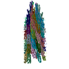
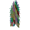
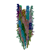
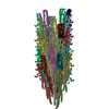
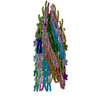
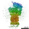
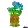
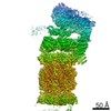
| Title | Cryo-EM structure of the wild-type flagellar filament of the Firmicute Kurthia | |||||||||||||||
 Components Components | Flagellin | |||||||||||||||
 Keywords Keywords | PROTEIN FIBRIL / Gram-Positive Bacteria Flagella / Helical / Wild-Type | |||||||||||||||
| Function / homology |  Function and homology information Function and homology informationbacterial-type flagellum / structural molecule activity / extracellular region Similarity search - Function | |||||||||||||||
| Biological species |  Kurthia sp. 11kri321 (bacteria) Kurthia sp. 11kri321 (bacteria) | |||||||||||||||
| Method | ELECTRON MICROSCOPY / helical reconstruction / cryo EM / Resolution: 2.8 Å | |||||||||||||||
 Authors Authors | Blum, T.B. / Abrahams, J.P. | |||||||||||||||
| Funding support |  Switzerland, 4items Switzerland, 4items
| |||||||||||||||
 Citation Citation | Journal: Sci Rep / Year: 2019 Title: The wild-type flagellar filament of the Firmicute Kurthia at 2.8 Å resolution in vivo. Authors: Thorsten B Blum / Sevasti Filippidou / Mathilda Fatton / Pilar Junier / Jan Pieter Abrahams /  Abstract: Bacteria swim and swarm by rotating the micrometers long, helical filaments of their flagella. They change direction by reversing their flagellar rotation, which switches the handedness of the ...Bacteria swim and swarm by rotating the micrometers long, helical filaments of their flagella. They change direction by reversing their flagellar rotation, which switches the handedness of the filament's supercoil. So far, all studied functional filaments are composed of a mixture of L- and R-state flagellin monomers. Here we show in a study of the wild type Firmicute Kurthia sp., that curved, functional filaments can adopt a conformation in vivo that is closely related to a uniform, all-L-state. This sheds additional light on transitions of the flagellar supercoil and uniquely reveals the atomic structure of a wild-type flagellar filament in vivo, including six residues showing clearly densities of O-linked glycosylation. | |||||||||||||||
| History |
|
- Structure visualization
| Movie |
Movie viewer |
|---|---|
| Structure viewer | Molecule: MolmilJmol/JSmol |
- Downloads & links
-Download
| PDBx/mmCIF format | 6t17.cif.gz | 3.5 MB | Display | PDBx/mmCIF format |
|---|---|---|---|---|
| PDB format | pdb6t17.ent.gz | Display | PDB format | |
| PDBx/mmJSON format | 6t17.json.gz | Tree view | PDBx/mmJSON format | |
| Others |  Other downloads Other downloads |
-Validation report
| Arichive directory | https://data.pdbj.org/pub/pdb/validation_reports/t1/6t17ftp://data.pdbj.org/pub/pdb/validation_reports/t1/6t17 | HTTPS FTP |
|---|
-Related structure data
| Related structure data | 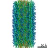 10362MC M: map data used to model this data C: citing same article ( |
|---|---|
| Similar structure data |
-Links
 PDBj
PDBj- Assembly
| Deposited unit | 
|
|---|---|
| 1 |
|
-Components
| #1: Protein | Mass: 29530.055 Da / Num. of mol.: 44 Source method: isolated from a genetically manipulated source Source: (gene. exp.) Kurthia sp. 11kri321 (bacteria) / Gene: ASO14_2420 / Production host: Kurthia sp. 11kri321 (bacteria) / References: UniProt: A0A0X9VHV9 |
|---|
-Experimental details
-Experiment
| Experiment | Method: ELECTRON MICROSCOPY |
|---|---|
| EM experiment | Aggregation state: FILAMENT / 3D reconstruction method: helical reconstruction |
- Sample preparation
| Component | Name: Flagellar Filament / Type: COMPLEX / Entity ID: all / Source: NATURAL |
|---|---|
| Source (natural) | Organism: Kurthia sp. 11kri321 (bacteria) |
| Buffer solution | pH: 7.3 |
| Specimen | Embedding applied: NO / Shadowing applied: NO / Staining applied: NO / Vitrification applied: YES |
| Vitrification | Instrument: LEICA EM GP / Cryogen name: ETHANE |
- Electron microscopy imaging
| Experimental equipment |  Model: Titan Krios / Image courtesy: FEI Company |
|---|---|
| Microscopy | Model: FEI TITAN KRIOS |
| Electron gun | Electron source:  FIELD EMISSION GUN / Accelerating voltage: 300 kV / Illumination mode: FLOOD BEAM FIELD EMISSION GUN / Accelerating voltage: 300 kV / Illumination mode: FLOOD BEAM |
| Electron lens | Mode: BRIGHT FIELD |
| Image recording | Electron dose: 86 e/Å2 / Detector mode: SUPER-RESOLUTION / Film or detector model: GATAN K2 SUMMIT (4k x 4k) |
- Processing
| EM software | Name: SerialEM / Category: image acquisition |
|---|---|
| CTF correction | Type: PHASE FLIPPING AND AMPLITUDE CORRECTION |
| Helical symmerty | Angular rotation/subunit: 65.45 ° / Axial rise/subunit: 4.83 Å / Axial symmetry: C1 |
| 3D reconstruction | Resolution: 2.8 Å / Resolution method: FSC 0.143 CUT-OFF / Num. of particles: 9270 / Algorithm: BACK PROJECTION / Num. of class averages: 1 / Symmetry type: HELICAL |
