 ムービー
ムービー コントローラー


+ データを開く

- 基本情報
基本情報
| 登録情報 | データベース: EMDB / ID: EMD-10211 | |||||||||
|---|---|---|---|---|---|---|---|---|---|---|
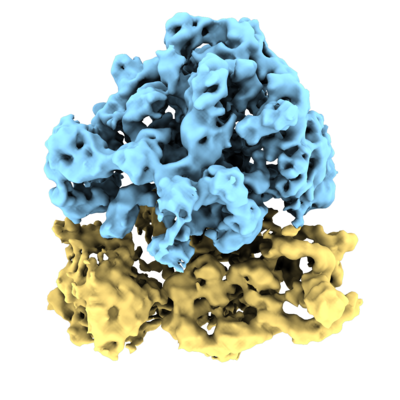
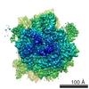
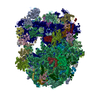
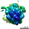
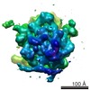
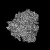
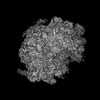
| タイトル | Subtomogram average of E. coli 70S ribosome from FISE tilt series | |||||||||
 マップデータ マップデータ | ||||||||||
 試料 試料 |
| |||||||||
| 生物種 |  | |||||||||
| 手法 | サブトモグラム平均法 / クライオ電子顕微鏡法 / 解像度: 9.0 Å | |||||||||
 データ登録者 データ登録者 | Eisenstein F / Danev R / Pilhofer M | |||||||||
 引用 引用 | ジャーナル: J Struct Biol / 年: 2019 タイトル: Improved applicability and robustness of fast cryo-electron tomography data acquisition. 著者: Fabian Eisenstein / Radostin Danev / Martin Pilhofer /   要旨: The power of cryo-electron tomography (cryoET) lies in its capability to characterize macromolecules in their cellular context. Structure determination by cryoET, however, is time-consuming compared ...The power of cryo-electron tomography (cryoET) lies in its capability to characterize macromolecules in their cellular context. Structure determination by cryoET, however, is time-consuming compared to single particle approaches. A recent study reported significant acceleration of data acquisition by a fast-incremental single-exposure (FISE) tilt series scheme. Here we improved the method and evaluated its efficiency and performance. We show that (1) FISE combined with the latest generation of direct electron detectors speeds up collection considerably, (2) previous generation (pre-2017) double-tilt axis Titan Krios holders are also suitable for FISE data acquisition, (3) x, y and z-specimen shifts can be compensated for, and (4) FISE tilt series data can generate averages of sub-nanometer resolution. These advances will allow for a widespread adoption of cryoET for high-throughput in situ studies and high-resolution structure determination across different biological research disciplines. #2: ジャーナル: J.Struct.Biol. / 年: 2019タイトル: Improved applicability and robustness of fast cryo-electron tomography data acquisition 著者: Eisenstein F / Danev R / Pilhofer M | |||||||||
| 履歴 |
|
- 構造の表示
構造の表示
| ムービー |
 ムービービューア ムービービューア |
|---|---|
| 構造ビューア | EMマップ: SurfViewMolmilJmol/JSmol |
| 添付画像 |
- ダウンロードとリンク
-EMDBアーカイブ
| マップデータ | emd_10211.map.gz | 5.5 MB | EMDBマップデータ形式 | |
|---|---|---|---|---|
| ヘッダ (付随情報) | emd-10211-v30.xmlemd-10211.xml | 11.5 KB 11.5 KB | 表示 表示 | EMDBヘッダ |
| 画像 |  emd_10211.png emd_10211.png | 167.8 KB | ||
| アーカイブディレクトリ |  http://ftp.pdbj.org/pub/emdb/structures/EMD-10211ftp://ftp.pdbj.org/pub/emdb/structures/EMD-10211 http://ftp.pdbj.org/pub/emdb/structures/EMD-10211ftp://ftp.pdbj.org/pub/emdb/structures/EMD-10211 | HTTPS FTP |
-検証レポート
| 文書・要旨 | emd_10211_validation.pdf.gz | 224.1 KB | 表示 | EMDB検証レポート |
|---|---|---|---|---|
| 文書・詳細版 | emd_10211_full_validation.pdf.gz | 223.3 KB | 表示 | |
| XML形式データ | emd_10211_validation.xml.gz | 6.2 KB | 表示 | |
| アーカイブディレクトリ | https://ftp.pdbj.org/pub/emdb/validation_reports/EMD-10211ftp://ftp.pdbj.org/pub/emdb/validation_reports/EMD-10211 | HTTPS FTP |
-関連構造データ
| 類似構造データ | |
|---|---|
| 電子顕微鏡画像生データ | EMPIAR-10304 (タイトル: Improved applicability and robustness of fast cryo-electron tomography data acquisition Data size: 21.6 / Data #1: FISEtiltseries [tilt series]) |

-リンク
| EMDBのページ | EMDB (EBI/PDBe) / EMDataResource |
|---|---|
| 「今月の分子」の関連する項目 |

-マップ
| ファイル | ダウンロード / ファイル: emd_10211.map.gz / 形式: CCP4 / 大きさ: 48.9 MB / タイプ: IMAGE STORED AS FLOATING POINT NUMBER (4 BYTES) | ||||||||||||||||||||||||||||||||||||||||||||||||||||||||||||
|---|---|---|---|---|---|---|---|---|---|---|---|---|---|---|---|---|---|---|---|---|---|---|---|---|---|---|---|---|---|---|---|---|---|---|---|---|---|---|---|---|---|---|---|---|---|---|---|---|---|---|---|---|---|---|---|---|---|---|---|---|---|
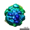
| 投影像・断面図 | 画像のコントロール
画像は Spider により作成 | ||||||||||||||||||||||||||||||||||||||||||||||||||||||||||||
| ボクセルのサイズ | X=Y=Z: 2.1 Å | ||||||||||||||||||||||||||||||||||||||||||||||||||||||||||||
| 密度 |
| ||||||||||||||||||||||||||||||||||||||||||||||||||||||||||||
| 対称性 | 空間群: 1 | ||||||||||||||||||||||||||||||||||||||||||||||||||||||||||||
| 詳細 | EMDB XML:
CCP4マップ ヘッダ情報:
| ||||||||||||||||||||||||||||||||||||||||||||||||||||||||||||
 Z (Sec.)
 Y (Row.)
 X (Col.)


-添付データ
- 試料の構成要素
-全体 : 70S ribosome
| 全体 | 名称: 70S ribosome |
|---|---|
| 要素 |
|
-超分子 #1: 70S ribosome
| 超分子 | 名称: 70S ribosome / タイプ: complex / ID: 1 / 親要素: 0 |
|---|---|
| 由来(天然) | 生物種: |
-実験情報
-構造解析
| 手法 | クライオ電子顕微鏡法 |
|---|---|
 解析 解析 | サブトモグラム平均法 |
| 試料の集合状態 | particle |
-試料調製
| 緩衝液 | pH: 7.5 |
|---|---|
| グリッド | モデル: Quantifoil R2/2 / 材質: COPPER / メッシュ: 200 |
| 凍結 | 凍結剤: ETHANE / チャンバー内湿度: 100 % |
- 電子顕微鏡法
| 顕微鏡 | FEI TITAN KRIOS |
|---|---|
| 詳細 | single-tilt axis holder |
| 撮影 | フィルム・検出器のモデル: GATAN K3 BIOQUANTUM (6k x 4k) 平均露光時間: 0.5 sec. / 平均電子線量: 150.0 e/Å2 / 詳細: FISEtomo SerialEM script |
| 電子線 | 加速電圧: 300 kV / 電子線源:  FIELD EMISSION GUN FIELD EMISSION GUN |
| 電子光学系 | 照射モード: FLOOD BEAM / 撮影モード: BRIGHT FIELD / Cs: 2.7 mm |
| 試料ステージ | 試料ホルダーモデル: FEI TITAN KRIOS AUTOGRID HOLDER ホルダー冷却材: NITROGEN |
| 実験機器 |  モデル: Titan Krios / 画像提供: FEI Company |
