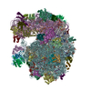
 Movie
Movie Controller

[English] 日本語
 Yorodumi
- EMDB-5041: Ribosome structure : Structural survey of large protein complexes... -
+ Open data

- Basic information
| Entry | Database: EMDB / ID: EMD-5041 | |||||||||
|---|---|---|---|---|---|---|---|---|---|---|
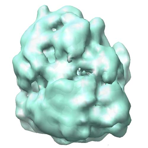
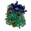
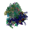
| Title | Ribosome structure : Structural survey of large protein complexes in Desulfovibrio vulgaris Hildenborough (DvH) | |||||||||
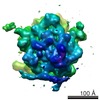
 Map data Map data | Ribosome structure from Desulfovibrio vulgaris Hildenborough (DvH) | |||||||||
 Sample Sample |
| |||||||||
 Keywords Keywords | Ribosome / Desulfovibrio vulgaris / DvH | |||||||||
| Biological species |  Desulfovibrio vulgaris (bacteria) Desulfovibrio vulgaris (bacteria) | |||||||||
| Method | single particle reconstruction / negative staining / Resolution: 24.0 Å | |||||||||
 Authors Authors | Han B-G / Dong M / Liu H / Camp L / Geller J / Singer M / Hazen TC / Choi M / Witkowska HE / Ball DA ...Han B-G / Dong M / Liu H / Camp L / Geller J / Singer M / Hazen TC / Choi M / Witkowska HE / Ball DA / Typke D / Downing KH / Shatsky M / Brenner SE / Chandonia J-M / Biggin MD / Glaeser RM | |||||||||
 Citation Citation | Journal: Proc Natl Acad Sci U S A / Year: 2009 Title: Survey of large protein complexes in D. vulgaris reveals great structural diversity. Authors: Bong-Gyoon Han / Ming Dong / Haichuan Liu / Lauren Camp / Jil Geller / Mary Singer / Terry C Hazen / Megan Choi / H Ewa Witkowska / David A Ball / Dieter Typke / Kenneth H Downing / Maxim ...Authors: Bong-Gyoon Han / Ming Dong / Haichuan Liu / Lauren Camp / Jil Geller / Mary Singer / Terry C Hazen / Megan Choi / H Ewa Witkowska / David A Ball / Dieter Typke / Kenneth H Downing / Maxim Shatsky / Steven E Brenner / John-Marc Chandonia / Mark D Biggin / Robert M Glaeser /  Abstract: An unbiased survey has been made of the stable, most abundant multi-protein complexes in Desulfovibrio vulgaris Hildenborough (DvH) that are larger than Mr approximately 400 k. The quaternary ...An unbiased survey has been made of the stable, most abundant multi-protein complexes in Desulfovibrio vulgaris Hildenborough (DvH) that are larger than Mr approximately 400 k. The quaternary structures for 8 of the 16 complexes purified during this work were determined by single-particle reconstruction of negatively stained specimens, a success rate approximately 10 times greater than that of previous "proteomic" screens. In addition, the subunit compositions and stoichiometries of the remaining complexes were determined by biochemical methods. Our data show that the structures of only two of these large complexes, out of the 13 in this set that have recognizable functions, can be modeled with confidence based on the structures of known homologs. These results indicate that there is significantly greater variability in the way that homologous prokaryotic macromolecular complexes are assembled than has generally been appreciated. As a consequence, we suggest that relying solely on previously determined quaternary structures for homologous proteins may not be sufficient to properly understand their role in another cell of interest. | |||||||||
| History |
|
- Structure visualization
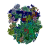
| Movie |
 Movie viewer Movie viewer |
|---|---|
| Structure viewer | EM map: SurfViewMolmilJmol/JSmol |
| Supplemental images |
- Downloads & links
-EMDB archive
| Map data | emd_5041.map.gz | 6.9 MB | EMDB map data format | |
|---|---|---|---|---|
| Header (meta data) | emd-5041-v30.xmlemd-5041.xml | 11.1 KB 11.1 KB | Display Display | EMDB header |
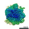
| Images |  emd_5041_1.jpg emd_5041_1.jpg | 44.9 KB | ||
| Archive directory |  http://ftp.pdbj.org/pub/emdb/structures/EMD-5041ftp://ftp.pdbj.org/pub/emdb/structures/EMD-5041 http://ftp.pdbj.org/pub/emdb/structures/EMD-5041ftp://ftp.pdbj.org/pub/emdb/structures/EMD-5041 | HTTPS FTP |
-Related structure data
| Related structure data | 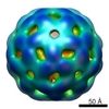 5042C 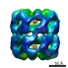 5043C 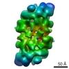 5044C 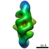 5045C  5046C  5047C C: citing same article ( |
|---|---|
| Similar structure data |


-Links
| EMDB pages | EMDB (EBI/PDBe) / EMDataResource |
|---|---|
| Related items in Molecule of the Month |


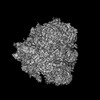
-Map
| File | Download / File: emd_5041.map.gz / Format: CCP4 / Size: 7.8 MB / Type: IMAGE STORED AS FLOATING POINT NUMBER (4 BYTES) | ||||||||||||||||||||||||||||||||||||||||||||||||||||||||||||||||||||
|---|---|---|---|---|---|---|---|---|---|---|---|---|---|---|---|---|---|---|---|---|---|---|---|---|---|---|---|---|---|---|---|---|---|---|---|---|---|---|---|---|---|---|---|---|---|---|---|---|---|---|---|---|---|---|---|---|---|---|---|---|---|---|---|---|---|---|---|---|---|
| Annotation | Ribosome structure from Desulfovibrio vulgaris Hildenborough (DvH) | ||||||||||||||||||||||||||||||||||||||||||||||||||||||||||||||||||||
| Projections & slices | Image control
Images are generated by Spider. | ||||||||||||||||||||||||||||||||||||||||||||||||||||||||||||||||||||
| Voxel size | X=Y=Z: 3.175 Å | ||||||||||||||||||||||||||||||||||||||||||||||||||||||||||||||||||||
| Density |
| ||||||||||||||||||||||||||||||||||||||||||||||||||||||||||||||||||||
| Symmetry | Space group: 1 | ||||||||||||||||||||||||||||||||||||||||||||||||||||||||||||||||||||
| Details | EMDB XML:
CCP4 map header:
| ||||||||||||||||||||||||||||||||||||||||||||||||||||||||||||||||||||
 Z (Sec.)
 Y (Row.)
 X (Col.)


-Supplemental data
- Sample components
-Entire : 70S ribosome from Desulfovibrio vulgaris Hildenborough
| Entire | Name: 70S ribosome from Desulfovibrio vulgaris Hildenborough |
|---|---|
| Components |
|
-Supramolecule #1000: 70S ribosome from Desulfovibrio vulgaris Hildenborough
| Supramolecule | Name: 70S ribosome from Desulfovibrio vulgaris Hildenborough type: sample / ID: 1000 / Oligomeric state: monomer / Number unique components: 1 |
|---|---|
| Molecular weight | Experimental: 3 MDa / Theoretical: 3 MDa / Method: Sedimentaion |
-Supramolecule #1: Ribosome
| Supramolecule | Name: Ribosome / type: complex / ID: 1 / Recombinant expression: No / Database: NCBI / Ribosome-details: ribosome-prokaryote: ALL |
|---|---|
| Source (natural) | Organism: Desulfovibrio vulgaris (bacteria) / Strain: Hildenborough / synonym: DvH |
| Molecular weight | Experimental: 3 MDa / Theoretical: 3 MDa |
-Experimental details
-Structure determination
| Method | negative staining |
|---|---|
 Processing Processing | single particle reconstruction |
| Aggregation state | particle |
-Sample preparation
| Concentration | 0.03 mg/mL |
|---|---|
| Buffer | pH: 7.5 Details: 20mM Tris buffer with 60mM NH4Cl, 6mM MgCl2, 0.5mM EDTA and 1mM DTT |
| Staining | Type: NEGATIVE Details: Three microliter of 2% w/v uranyl acetate stain was applied to the EM grid for 1 min. |
| Grid | Details: carbon-coated and glow-discharged 300 mesh copper grid |
| Vitrification | Cryogen name: NONE / Instrument: OTHER |
- Electron microscopy
| Microscope | JEOL 4000EX |
|---|---|
| Temperature | Average: 293 K |
| Alignment procedure | Legacy - Astigmatism: objective lens astigmatism was corrected at 60,000 times magnification |
| Date | Jul 8, 2008 |
| Image recording | Category: FILM / Film or detector model: KODAK SO-163 FILM / Digitization - Scanner: NIKON SUPER COOLSCAN 9000 / Digitization - Sampling interval: 6.35 µm / Number real images: 85 / Average electron dose: 17 e/Å2 Details: The images were scanned with a resolution of 6.35 micro m per pixel and later averaged 2 fold in each direction. Bits/pixel: 8 |
| Tilt angle min | 0 |
| Tilt angle max | 0 |
| Electron beam | Acceleration voltage: 400 kV / Electron source:  FIELD EMISSION GUN FIELD EMISSION GUN |
| Electron optics | Calibrated magnification: 40000 / Illumination mode: FLOOD BEAM / Imaging mode: BRIGHT FIELD / Cs: 4.1 mm / Nominal defocus max: 2.0 µm / Nominal defocus min: 0.6 µm / Nominal magnification: 40000 |
| Sample stage | Specimen holder: Side entry room T / Specimen holder model: OTHER |
-Image processing
| CTF correction | Details: Each particle |
|---|---|
| Final reconstruction | Algorithm: OTHER / Resolution.type: BY AUTHOR / Resolution: 24.0 Å / Resolution method: FSC 0.5 CUT-OFF / Software - Name: SPIDER / Number images used: 11000 |
