 Movie
Movie Controller

[English] 日本語
 Yorodumi
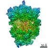
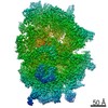
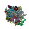
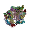
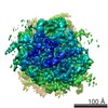
- EMDB-10179: mt-SSU late assembly intermediate of wild-type Trypanosoma brucei... -
+ Open data

- Basic information
| Entry | Database: EMDB / ID: EMD-10179 | |||||||||
|---|---|---|---|---|---|---|---|---|---|---|
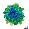
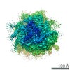
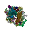
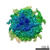
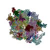
| Title | mt-SSU late assembly intermediate of wild-type Trypanosoma brucei brucei | |||||||||
 Map data Map data | mt-SSU late assembly intermediate of wild-type Trypanosoma brucei brucei | |||||||||
 Sample Sample |
| |||||||||
| Biological species |  | |||||||||
| Method | single particle reconstruction / cryo EM / Resolution: 5.9 Å | |||||||||
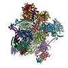
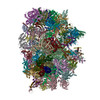
 Authors Authors | Saurer M / Ramrath D / Niemann M / Calderaro S / Prange C / Mattei S / Scaiola A / Leitner A / Bieri P / Horn EK ...Saurer M / Ramrath D / Niemann M / Calderaro S / Prange C / Mattei S / Scaiola A / Leitner A / Bieri P / Horn EK / Leibundgut M / Schneider A / Boehringer D / Ban N | |||||||||
| Funding support |  Switzerland, 1 items Switzerland, 1 items
| |||||||||
 Citation Citation | Journal: Science / Year: 2019 Title: Mitoribosomal small subunit biogenesis in trypanosomes involves an extensive assembly machinery. Authors: Martin Saurer / David J F Ramrath / Moritz Niemann / Salvatore Calderaro / Céline Prange / Simone Mattei / Alain Scaiola / Alexander Leitner / Philipp Bieri / Elke K Horn / Marc Leibundgut ...Authors: Martin Saurer / David J F Ramrath / Moritz Niemann / Salvatore Calderaro / Céline Prange / Simone Mattei / Alain Scaiola / Alexander Leitner / Philipp Bieri / Elke K Horn / Marc Leibundgut / Daniel Boehringer / André Schneider / Nenad Ban / Abstract: Mitochondrial ribosomes (mitoribosomes) are large ribonucleoprotein complexes that synthesize proteins encoded by the mitochondrial genome. An extensive cellular machinery responsible for ribosome ...Mitochondrial ribosomes (mitoribosomes) are large ribonucleoprotein complexes that synthesize proteins encoded by the mitochondrial genome. An extensive cellular machinery responsible for ribosome assembly has been described only for eukaryotic cytosolic ribosomes. Here we report that the assembly of the small mitoribosomal subunit in involves a large number of factors and proceeds through the formation of assembly intermediates, which we analyzed by using cryo-electron microscopy. One of them is a 4-megadalton complex, referred to as the small subunit assemblosome, in which we identified 34 factors that interact with immature ribosomal RNA (rRNA) and recognize its functionally important regions. The assembly proceeds through large-scale conformational changes in rRNA coupled with successive incorporation of mitoribosomal proteins, providing an example for the complexity of the ribosomal assembly process in mitochondria. | |||||||||
| History |
|
- Structure visualization
| Movie |
 Movie viewer Movie viewer |
|---|---|
| Structure viewer | EM map: SurfViewMolmilJmol/JSmol |
| Supplemental images |

- Downloads & links
-EMDB archive
| Map data | emd_10179.map.gz | 10.6 MB | EMDB map data format | |
|---|---|---|---|---|
| Header (meta data) | emd-10179-v30.xmlemd-10179.xml | 26.7 KB 26.7 KB | Display Display | EMDB header |
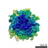
| Images | 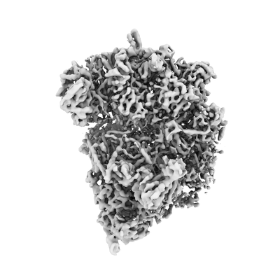 emd_10179.png emd_10179.png | 49.8 KB | ||
| Archive directory |  http://ftp.pdbj.org/pub/emdb/structures/EMD-10179ftp://ftp.pdbj.org/pub/emdb/structures/EMD-10179 http://ftp.pdbj.org/pub/emdb/structures/EMD-10179ftp://ftp.pdbj.org/pub/emdb/structures/EMD-10179 | HTTPS FTP |
-Related structure data

-Links
| EMDB pages | EMDB (EBI/PDBe) / EMDataResource |
|---|---|
| Related items in Molecule of the Month |

-Map
| File | Download / File: emd_10179.map.gz / Format: CCP4 / Size: 64 MB / Type: IMAGE STORED AS FLOATING POINT NUMBER (4 BYTES) | ||||||||||||||||||||||||||||||||||||||||||||||||||||||||||||
|---|---|---|---|---|---|---|---|---|---|---|---|---|---|---|---|---|---|---|---|---|---|---|---|---|---|---|---|---|---|---|---|---|---|---|---|---|---|---|---|---|---|---|---|---|---|---|---|---|---|---|---|---|---|---|---|---|---|---|---|---|---|
| Annotation | mt-SSU late assembly intermediate of wild-type Trypanosoma brucei brucei | ||||||||||||||||||||||||||||||||||||||||||||||||||||||||||||
| Projections & slices | Image control
Images are generated by Spider. | ||||||||||||||||||||||||||||||||||||||||||||||||||||||||||||
| Voxel size | X=Y=Z: 1.69531 Å | ||||||||||||||||||||||||||||||||||||||||||||||||||||||||||||
| Density |
| ||||||||||||||||||||||||||||||||||||||||||||||||||||||||||||
| Symmetry | Space group: 1 | ||||||||||||||||||||||||||||||||||||||||||||||||||||||||||||
| Details | EMDB XML:
CCP4 map header:
| ||||||||||||||||||||||||||||||||||||||||||||||||||||||||||||
 Z (Sec.)
 Y (Row.)
 X (Col.)


-Supplemental data
- Sample components
-Entire : wild-type mt-SSU late assembly intermediate
| Entire | Name: wild-type mt-SSU late assembly intermediate |
|---|---|
| Components |
|
-Supramolecule #1: wild-type mt-SSU late assembly intermediate
| Supramolecule | Name: wild-type mt-SSU late assembly intermediate / type: complex / ID: 1 / Parent: 0 / Macromolecule list: #1-#103 |
|---|---|
| Source (natural) | Organism: |
-Experimental details
-Structure determination
| Method | cryo EM |
|---|---|
 Processing Processing | single particle reconstruction |
| Aggregation state | particle |
-Sample preparation
| Buffer | pH: 7.4 |
|---|---|
| Grid | Model: Quantifoil R2/2 / Material: COPPER / Mesh: 300 / Support film - Material: CARBON / Support film - topology: CONTINUOUS / Pretreatment - Type: GLOW DISCHARGE / Pretreatment - Atmosphere: AIR / Details: 15 mA |
| Vitrification | Cryogen name: ETHANE-PROPANE / Chamber humidity: 100 % / Chamber temperature: 277 K / Instrument: FEI VITROBOT MARK IV |
| Details | Mitochondrial ribosomal small subunit middle assembly intermediate of wild-type Trypanosoma brucei brucei Lister strain 427 |
- Electron microscopy
| Microscope | FEI TITAN KRIOS |
|---|---|
| Image recording | Film or detector model: FEI FALCON III (4k x 4k) / Detector mode: INTEGRATING / Digitization - Sampling interval: 14.0 µm / Number grids imaged: 1 / Number real images: 13503 / Average exposure time: 1.5 sec. / Average electron dose: 75.0 e/Å2 Details: Images were collected in movie-mode at 40 frames per second |
| Electron beam | Acceleration voltage: 300 kV / Electron source:  FIELD EMISSION GUN FIELD EMISSION GUN |
| Electron optics | C2 aperture diameter: 100.0 µm / Calibrated defocus max: 4.5 µm / Calibrated defocus min: 1.0 µm / Calibrated magnification: 129032 / Illumination mode: FLOOD BEAM / Imaging mode: BRIGHT FIELD / Cs: 2.7 mm / Nominal magnification: 75000 |
| Sample stage | Specimen holder model: FEI TITAN KRIOS AUTOGRID HOLDER / Cooling holder cryogen: NITROGEN |
| Experimental equipment |  Model: Titan Krios / Image courtesy: FEI Company |
+Image processing
-Atomic model buiding 1
| Details | Starting models of ribosomal proteins of the T. brucei mitochondrial ribosomal small subunit were taken from PDB 6HIW. Mitchondrial ribosomal small subunit assembly factors were built de novo. |
|---|---|
| Refinement | Protocol: OTHER |
