ムービー
ムービー コントローラー


+ データを開く

- 基本情報
基本情報
| 登録情報 | データベース: EMDB / ID: EMD-10091 | |||||||||
|---|---|---|---|---|---|---|---|---|---|---|
| タイトル | Granulovirus occlusion bodies by serial electron diffraction | |||||||||
 マップデータ マップデータ | ||||||||||
 試料 試料 |
| |||||||||
 キーワード キーワード | granulovirus / occlusion body / serial crystallography / viral protein | |||||||||
| 機能・相同性 | Polyhedrin / Polyhedrin / viral occlusion body / structural molecule activity / Granulin 機能・相同性情報 機能・相同性情報 | |||||||||
| 生物種 |  Cydia pomonella granulosis virus (isolate Mexico/1963) (ウイルス) / Cydia pomonella granulosis virus (isolate Mexican) (ウイルス) Cydia pomonella granulosis virus (isolate Mexico/1963) (ウイルス) / Cydia pomonella granulosis virus (isolate Mexican) (ウイルス) | |||||||||
| 手法 | 電子線結晶学 / クライオ電子顕微鏡法 / 解像度: 1.6 Å | |||||||||
 データ登録者 データ登録者 | Buecker R / Mehrabi P | |||||||||
 引用 引用 | ジャーナル: Nat Commun / 年: 2020 タイトル: Serial protein crystallography in an electron microscope. 著者: Robert Bücker / Pascal Hogan-Lamarre / Pedram Mehrabi / Eike C Schulz / Lindsey A Bultema / Yaroslav Gevorkov / Wolfgang Brehm / Oleksandr Yefanov / Dominik Oberthür / Günther H Kassier / R J Dwayne Miller /   要旨: Serial X-ray crystallography at free-electron lasers allows to solve biomolecular structures from sub-micron-sized crystals. However, beam time at these facilities is scarce, and involved sample ...Serial X-ray crystallography at free-electron lasers allows to solve biomolecular structures from sub-micron-sized crystals. However, beam time at these facilities is scarce, and involved sample delivery techniques are required. On the other hand, rotation electron diffraction (MicroED) has shown great potential as an alternative means for protein nano-crystallography. Here, we present a method for serial electron diffraction of protein nanocrystals combining the benefits of both approaches. In a scanning transmission electron microscope, crystals randomly dispersed on a sample grid are automatically mapped, and a diffraction pattern at fixed orientation is recorded from each at a high acquisition rate. Dose fractionation ensures minimal radiation damage effects. We demonstrate the method by solving the structure of granulovirus occlusion bodies and lysozyme to resolutions of 1.55 Å and 1.80 Å, respectively. Our method promises to provide rapid structure determination for many classes of materials with minimal sample consumption, using readily available instrumentation. | |||||||||
| 履歴 |
|
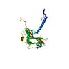
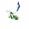
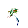
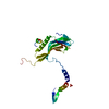
- 構造の表示
構造の表示
| ムービー |
ムービービューア |
|---|---|
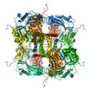
| 構造ビューア | EMマップ: SurfViewMolmilJmol/JSmol |
| 添付画像 |

- ダウンロードとリンク
-EMDBアーカイブ
| マップデータ | emd_10091.map.gz | 2.2 MB | EMDBマップデータ形式 | |
|---|---|---|---|---|
| ヘッダ (付随情報) | emd-10091-v30.xmlemd-10091.xml | 12.8 KB 12.8 KB | 表示 表示 | EMDBヘッダ |
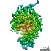
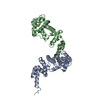
| 画像 | 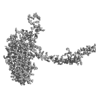 emd_10091.png emd_10091.png | 104.9 KB | ||
| Filedesc metadata | emd-10091.cif.gz | 5.5 KB | ||
| Filedesc structureFactors | emd_10091_sf.cif.gz | 687.9 KB | ||
| アーカイブディレクトリ |  http://ftp.pdbj.org/pub/emdb/structures/EMD-10091ftp://ftp.pdbj.org/pub/emdb/structures/EMD-10091 http://ftp.pdbj.org/pub/emdb/structures/EMD-10091ftp://ftp.pdbj.org/pub/emdb/structures/EMD-10091 | HTTPS FTP |
-検証レポート
| 文書・要旨 | emd_10091_validation.pdf.gz | 382.6 KB | 表示 | EMDB検証レポート |
|---|---|---|---|---|
| 文書・詳細版 | emd_10091_full_validation.pdf.gz | 382.1 KB | 表示 | |
| XML形式データ | emd_10091_validation.xml.gz | 4.4 KB | 表示 | |
| CIF形式データ | emd_10091_validation.cif.gz | 5 KB | 表示 | |
| アーカイブディレクトリ | https://ftp.pdbj.org/pub/emdb/validation_reports/EMD-10091ftp://ftp.pdbj.org/pub/emdb/validation_reports/EMD-10091 | HTTPS FTP |
-関連構造データ


-リンク
| EMDBのページ | EMDB (EBI/PDBe) / EMDataResource |
|---|
-マップ
| ファイル | ダウンロード / ファイル: emd_10091.map.gz / 形式: CCP4 / 大きさ: 41.4 MB / タイプ: IMAGE STORED AS FLOATING POINT NUMBER (4 BYTES) | ||||||||||||||||||||||||||||||||||||||||||||||||||||||||||||
|---|---|---|---|---|---|---|---|---|---|---|---|---|---|---|---|---|---|---|---|---|---|---|---|---|---|---|---|---|---|---|---|---|---|---|---|---|---|---|---|---|---|---|---|---|---|---|---|---|---|---|---|---|---|---|---|---|---|---|---|---|---|
| 投影像・断面図 | 画像のコントロール
画像は Spider により作成 これらの図は立方格子座標系で作成されたものです | ||||||||||||||||||||||||||||||||||||||||||||||||||||||||||||
| ボクセルのサイズ | X=Y=Z: 0.3822 Å | ||||||||||||||||||||||||||||||||||||||||||||||||||||||||||||
| 密度 |
| ||||||||||||||||||||||||||||||||||||||||||||||||||||||||||||
| 対称性 | 空間群: 1 | ||||||||||||||||||||||||||||||||||||||||||||||||||||||||||||
| 詳細 | EMDB XML:
CCP4マップ ヘッダ情報:
| ||||||||||||||||||||||||||||||||||||||||||||||||||||||||||||
 Z (Sec.)
 Y (Row.)
 X (Col.)


-添付データ
- 試料の構成要素
-全体 : Cydia pomonella granulosis virus (isolate Mexican)
| 全体 | 名称: Cydia pomonella granulosis virus (isolate Mexican) (ウイルス) |
|---|---|
| 要素 |
|
-超分子 #1: Cydia pomonella granulosis virus (isolate Mexican)
| 超分子 | 名称: Cydia pomonella granulosis virus (isolate Mexican) / タイプ: virus / ID: 1 / 親要素: 0 / 含まれる分子: #1 / NCBI-ID: 654905 生物種: Cydia pomonella granulosis virus (isolate Mexican) ウイルスタイプ: VIRION / ウイルス・単離状態: OTHER / ウイルス・エンベロープ: No / ウイルス・中空状態: No |
|---|
-分子 #1: Granulin
| 分子 | 名称: Granulin / タイプ: protein_or_peptide / ID: 1 / コピー数: 1 / 光学異性体: LEVO |
|---|---|
| 由来(天然) | 生物種: Cydia pomonella granulosis virus (isolate Mexico/1963) (ウイルス) 株: isolate Mexico/1963 |
| 分子量 | 理論値: 29.378559 KDa |
| 組換発現 | 生物種: Cydia pomonella granulosis virus (isolate Mexican) (ウイルス) |
| 配列 | 文字列: MGYNKSLRYS RHDGTSCVID NHHLKSLGAV LNDVRRKKDR IREAEYEPII DIADQYMVTE DPFRGPGKNV RITLFKEIRR VHPDTMKLV CNWSGKEFLR ETWTRFISEE FPITTDQEIM DLWFELQLRP MHPNRCYKFT MQYALGAHPD YVAHDVIRQQ D PYYVGPNN ...文字列: MGYNKSLRYS RHDGTSCVID NHHLKSLGAV LNDVRRKKDR IREAEYEPII DIADQYMVTE DPFRGPGKNV RITLFKEIRR VHPDTMKLV CNWSGKEFLR ETWTRFISEE FPITTDQEIM DLWFELQLRP MHPNRCYKFT MQYALGAHPD YVAHDVIRQQ D PYYVGPNN IERINLSKKG FAFPLTCLQS VYNDNFERFF DDVLWPYFYR PLVYVGTTSA EIEEIMIEVS LLFKIKEFAP DV PLFTGPA Y UniProtKB: Granulin |
-分子 #2: water
| 分子 | 名称: water / タイプ: ligand / ID: 2 / コピー数: 90 / 式: HOH |
|---|---|
| 分子量 | 理論値: 18.015 Da |
| Chemical component information |  ChemComp-HOH: |
-実験情報
-構造解析
| 手法 | クライオ電子顕微鏡法 |
|---|---|
 解析 解析 | 電子線結晶学 |
| 試料の集合状態 | 3D array |
-試料調製
| 緩衝液 | pH: 7 |
|---|---|
| 凍結 | 凍結剤: ETHANE |
- 電子顕微鏡法
| 顕微鏡 | FEI TECNAI F20 |
|---|---|
| 温度 | 最低: 93.0 K / 最高: 100.0 K |
| 撮影 | フィルム・検出器のモデル: OTHER / デジタル化 - サイズ - 横: 1536 pixel / デジタル化 - サイズ - 縦: 512 pixel / 撮影したグリッド数: 1 / 回折像の数: 32000 / 平均露光時間: 0.01 sec. / 平均電子線量: 4.7 e/Å2 |
| 電子線 | 加速電圧: 200 kV / 電子線源:  FIELD EMISSION GUN FIELD EMISSION GUN |
| 電子光学系 | C2レンズ絞り径: 5.0 µm / 照射モード: OTHER / 撮影モード: DIFFRACTION / カメラ長: 2580 mm |
| 試料ステージ | 試料ホルダーモデル: GATAN 626 SINGLE TILT LIQUID NITROGEN CRYO TRANSFER HOLDER ホルダー冷却材: NITROGEN / 傾斜角度: 0 |
| 実験機器 |  モデル: Tecnai F20 / 画像提供: FEI Company |
-画像解析
| 最終 再構成 | 解像度のタイプ: BY AUTHOR / 解像度: 1.6 Å / 解像度の算出法: DIFFRACTION PATTERN/LAYERLINES / ソフトウェア - 名称: Coot |
|---|---|
| Molecular replacement | ソフトウェア - 名称: PHENIX (ver. 1.17) |
| Merging software list | ソフトウェア - 詳細: CrystFEL |
| Crystallography statistics | Number intensities measured: 9650574 / Number structure factors: 23422 / Fourier space coverage: 100 / R sym: 0.093 / R merge: 0.093 / Overall phase error: 13.64 / Overall phase residual: 1 / Phase error rejection criteria: 0 / High resolution: 1.6 Å / 殻 - Shell ID: 1 / 殻 - High resolution: 10.0 Å / 殻 - Low resolution: 1.55 Å / 殻 - Number structure factors: 1 / 殻 - Phase residual: 1 / 殻 - Fourier space coverage: 1 / 殻 - Multiplicity: 1 |