 Movie
Movie Controller

[English] 日本語
 Yorodumi
- PDB-6yng: MicroED structure of granulin determined from five native nanocry... -
+ Open data

- Basic information
| Entry | Database: PDB / ID: 6yng | ||||||
|---|---|---|---|---|---|---|---|
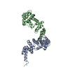
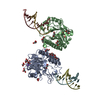
| Title | MicroED structure of granulin determined from five native nanocrystalline granulovirus occlusion bodies | ||||||
 Components Components | Granulin | ||||||
 Keywords Keywords | VIRAL PROTEIN / MicroED / nanocrystalography / native crystals | ||||||
| Function / homology | Polyhedrin / Polyhedrin / viral occlusion body / structural molecule activity / Granulin Function and homology information Function and homology information | ||||||
| Biological species |  Cydia pomonella granulosis virus Cydia pomonella granulosis virus | ||||||
| Method | ELECTRON CRYSTALLOGRAPHY / single particle reconstruction /  MOLECULAR REPLACEMENT / cryo EM / Resolution: 2.8 Å MOLECULAR REPLACEMENT / cryo EM / Resolution: 2.8 Å | ||||||
 Authors Authors | Bunker, R.D. | ||||||
 Citation Citation | Journal: To Be Published Title: MicroED structure of granulin determined from five native nanocrystalline granulovirus occlusion bodies Authors: Bunker, R.D. | ||||||
| History |
|
- Structure visualization
| Movie |
Movie viewer |
|---|---|
| Structure viewer | Molecule: MolmilJmol/JSmol |
- Downloads & links
-Download
| PDBx/mmCIF format | 6yng.cif.gz | 105.6 KB | Display | PDBx/mmCIF format |
|---|---|---|---|---|
| PDB format | pdb6yng.ent.gz | 82 KB | Display | PDB format |
| PDBx/mmJSON format | 6yng.json.gz | Tree view | PDBx/mmJSON format | |
| Others |  Other downloads Other downloads |
-Validation report
| Arichive directory | https://data.pdbj.org/pub/pdb/validation_reports/yn/6yngftp://data.pdbj.org/pub/pdb/validation_reports/yn/6yng | HTTPS FTP |
|---|
-Related structure data
| Related structure data |  5g0zS S: Starting model for refinement |
|---|---|
| Similar structure data |


-Links
 PDBj
PDBj- Assembly
| Deposited unit | 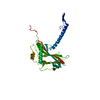
| ||||||||
|---|---|---|---|---|---|---|---|---|---|
| 1 | x 12
| ||||||||
| Unit cell |
|
-Components
| #1: Protein | Mass: 29378.559 Da / Num. of mol.: 1 / Source method: isolated from a natural source Source: (natural) Cydia pomonella granulosis virus (isolate Mexico/1963)Strain: isolate Mexico/1963 / References: UniProt: P87577 |
|---|
-Experimental details
-Experiment
| Experiment | Method: ELECTRON CRYSTALLOGRAPHY |
|---|---|
| EM experiment | Aggregation state: PARTICLE / 3D reconstruction method: single particle reconstruction |
- Sample preparation
| Component | Name: Cydia pomonella granulosis virus (isolate Mexican) / Type: VIRUS / Entity ID: all / Source: NATURAL |
|---|---|
| Molecular weight | Experimental value: NO |
| Source (natural) | Organism: Cydia pomonella granulosis virus (isolate Mexican) |
| Details of virus | Empty: NO / Enveloped: YES / Isolate: SPECIES / Type: VIRUS-LIKE PARTICLE |
| Natural host | Organism: Cydia pomonella |
| Buffer solution | pH: 7.4 |
| Specimen | Embedding applied: YES / Shadowing applied: NO / Staining applied: NO / Vitrification applied: YES |
| Specimen support | Grid material: COPPER / Grid mesh size: 200 divisions/in. / Grid type: Quantifoil R1.2/1.3 |
| EM embedding | Details: Undiluted sample was applied to glow-discharged 200 mesh copper Quantifoil. Material: Ice |
| Vitrification | Instrument: FEI VITROBOT MARK IV / Cryogen name: NITROGEN / Humidity: 100 % / Chamber temperature: 277 K / Details: 3 s blot, 0.5 s draining |
-Data collection
| Experimental equipment |  Model: Talos Arctica / Image courtesy: FEI Company |
|---|---|
| Microscopy | Model: FEI TALOS ARCTICA |
| Electron gun | Electron source: FIELD EMISSION GUN / Accelerating voltage: 200 kV / Illumination mode: FLOOD BEAM |
| Electron lens | Mode: BRIGHT FIELD / C2 aperture diameter: 20 µm |
| Image recording | Average exposure time: 2 sec. / Electron dose: 1 e/Å2 / Film or detector model: FEI CETA (4k x 4k) / Num. of diffraction images: 93 / Num. of grids imaged: 1 / Num. of real images: 93 Details: DIFFRACTON IMAGE RECORDED USING ROTATION METHOD DELPHI PHI 0.5 |
| EM diffraction | Camera length: 3600 mm |
| EM diffraction shell | Resolution: 2.8→30 Å / Fourier space coverage: 87.1 % / Multiplicity: 5 / Num. of structure factors: 4138 / Phase residual: 17.75 ° |
| EM diffraction stats | Details: 3D ELECTRON DIFFRACTION. RESOLUTION OF MERGED DATASET DEFINED BY AN INSCRIBED CIRCLE ON THE CAMERA SURFACE, WAS LIMITED TO 3.25 A BY THE CAMERA TO SAMPLE DISTANCE. Fourier space coverage: 87.1 % / High resolution: 2.8 Å / Num. of intensities measured: 20528 / Num. of structure factors: 4138 / Phase error rejection criteria: NULL / Rmerge: 0.321 |
- Processing
| Software | Name: PHENIX / Version: (dev_3313: ???) / Classification: refinement | ||||||||||||||||||||||||||||
|---|---|---|---|---|---|---|---|---|---|---|---|---|---|---|---|---|---|---|---|---|---|---|---|---|---|---|---|---|---|
| EM software |
| ||||||||||||||||||||||||||||
| Image processing | Details: DEVELOPMENT VERSION OF THE CETA-D CAMERA | ||||||||||||||||||||||||||||
| EM 3D crystal entity | ∠α: 90 ° / ∠β: 90 ° / ∠γ: 90 ° / A: 103.66 Å / B: 103.66 Å / C: 103.66 Å / Space group name: I23 / Space group num: 197 | ||||||||||||||||||||||||||||
| CTF correction | Type: NONE | ||||||||||||||||||||||||||||
| 3D reconstruction | Resolution: 2.8 Å / Resolution method: DIFFRACTION PATTERN/LAYERLINES Details: RESOLUTION OF MERGED DATASET DEFINED BY AN INSCRIBED CIRCLE ON THE CAMERA SURFACE, WAS LIMITED 3.0 TO BY THE CAMERA TO SAMPLE DISTANCE. DATA INTEGRATED WITH DIALS, COMBINED WITH POINTLESS, ...Details: RESOLUTION OF MERGED DATASET DEFINED BY AN INSCRIBED CIRCLE ON THE CAMERA SURFACE, WAS LIMITED 3.0 TO BY THE CAMERA TO SAMPLE DISTANCE. DATA INTEGRATED WITH DIALS, COMBINED WITH POINTLESS, AND SCALED WITH AIMLESS. INTENSITIES CONVERTED TO STRUCTURE FACTOR AMPLITUDES WITH STARANISO. INITIAL PHASES DETERMINED BY RIGID BODY FITTING A MODEL DERIVED FROM PDB ENTRY 5G0Z IN PHENIX.REFINE. FINAL REFINEMENT CARRIED OUT IN PHENIX.REFINE Symmetry type: 3D CRYSTAL | ||||||||||||||||||||||||||||
| Atomic model building | Space: RECIPROCAL | ||||||||||||||||||||||||||||
| Atomic model building | PDB-ID: 5G0Z Accession code: 5G0Z / Source name: PDB / Type: experimental model | ||||||||||||||||||||||||||||
| Refinement | Method to determine structure: MOLECULAR REPLACEMENT Starting model: 5G0Z Resolution: 2.8→2.8 Å / SU ML: 0.35 / Cross valid method: THROUGHOUT / σ(F): 1.39 / Phase error: 17.75
| ||||||||||||||||||||||||||||
| Solvent computation | Shrinkage radii: 0 Å / VDW probe radii: 0.4 Å | ||||||||||||||||||||||||||||
| Refine LS restraints |
| ||||||||||||||||||||||||||||
| LS refinement shell |
|
