 Movie
Movie Controller


+ Open data

- Basic information
| Entry | Database: PDB / ID: 3jvb | ||||||
|---|---|---|---|---|---|---|---|
| Title | Crystal structure of infectious baculovirus polyhedra | ||||||
 Components Components | Polyhedrin | ||||||
 Keywords Keywords | VIRAL PROTEIN / Jelly-roll / disulfide bond / domain swapping | ||||||
| Function / homology | Polyhedrin / Polyhedrin / viral occlusion body / structural molecule activity / Polyhedrin Function and homology information Function and homology information | ||||||
| Biological species |  Wiseana signata NPV (virus) Wiseana signata NPV (virus) | ||||||
| Method |  X-RAY DIFFRACTION / SYNCHROTRON / MOLECULAR REPLACEMENT / Resolution: 2.17 Å X-RAY DIFFRACTION / SYNCHROTRON / MOLECULAR REPLACEMENT / Resolution: 2.17 Å | ||||||
 Authors Authors | Coulibaly, F. / Chiu, E. / Metcalf, P. | ||||||
 Citation Citation | Journal: Proc.Natl.Acad.Sci.USA / Year: 2009 Title: The atomic structure of baculovirus polyhedra reveals the independent emergence of infectious crystals in DNA and RNA viruses Authors: Coulibaly, F. / Chiu, E. / Gutmann, S. / Rajendran, C. / Haebel, P.W. / Ikeda, K. / Mori, H. / Ward, V.K. / Schulze-Briese, C. / Metcalf, P. | ||||||
| History |
|
- Structure visualization
| Structure viewer | Molecule: MolmilJmol/JSmol |
|---|
- Downloads & links
-Download
| PDBx/mmCIF format | 3jvb.cif.gz | 56.9 KB | Display | PDBx/mmCIF format |
|---|---|---|---|---|
| PDB format | pdb3jvb.ent.gz | 40.6 KB | Display | PDB format |
| PDBx/mmJSON format | 3jvb.json.gz | Tree view | PDBx/mmJSON format | |
| Others |  Other downloads Other downloads |
-Validation report
| Arichive directory | https://data.pdbj.org/pub/pdb/validation_reports/jv/3jvbftp://data.pdbj.org/pub/pdb/validation_reports/jv/3jvb | HTTPS FTP |
|---|
-Related structure data
| Related structure data | 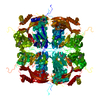 3jw6SC S: Starting model for refinement C: citing same article ( |
|---|---|
| Similar structure data |

-Links
 PDBj
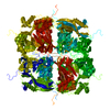
PDBj- Assembly
| Deposited unit | 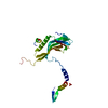
| |||||||||
|---|---|---|---|---|---|---|---|---|---|---|
| 1 | x 12
| |||||||||
| Unit cell |
| |||||||||
| Components on special symmetry positions |
| |||||||||
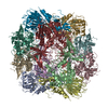
| Details | POLYHEDRA ARE VIRUS-CONTAINING CRYSTALS, WHICH REPRESENT THE MAIN INFECTIOUS FORM OF BACULOVIRUS. THE BIOLOGICAL ASSEMBLY IS THE WHOLE CRYSTAL. DODECAMERS OF THE POLYHEDRIN PROTEIN ARE PUTATIVE BUILDING BLOCKS OF THE CRYSTAL. INTERFACES INVOLVED IN GENERATING BIOMOLECULES 2-7 EXIST IN THE CRYSTAL BUT MAY NOT BE RELEVANT IN SOLUTION. |
-Components
| #1: Protein | Mass: 28705.818 Da / Num. of mol.: 1 / Source method: isolated from a natural source Details: Crystals were directly purified from insects (larvae of porina moths) naturally infected by a baculovirus (Wiseana spp. nucleopolyhedrosis virus) Source: (natural) Wiseana signata NPV (virus) / References: UniProt: O37157 |
|---|---|
| #2: Chemical | ChemComp-SO4 /   Mass: 96.063 Da / Num. of mol.: 1 / Source method: obtained synthetically / Formula: SO4 Mass: 96.063 Da / Num. of mol.: 1 / Source method: obtained synthetically / Formula: SO4 |
| #3: Water | ChemComp-HOH /  Mass: 18.015 Da / Num. of mol.: 86 / Source method: isolated from a natural source / Formula: H2O Mass: 18.015 Da / Num. of mol.: 86 / Source method: isolated from a natural source / Formula: H2O |
| Has protein modification | Y |
-Experimental details
-Experiment
| Experiment | Method: X-RAY DIFFRACTION / Number of used crystals: 19 |
|---|
- Sample preparation
| Crystal | Density Matthews: 1.560221 Å3/Da / Density % sol: 21.164995 % |
|---|---|
| Crystal grow | Temperature: 300 K / pH: 7 Details: Natural intracellular Crystals were directly purified from larvae of porina moths infected by a baculovirus (WNPV), pH 7, temperature 300K |
-Data collection
| Diffraction | Mean temperature: 100 K |
|---|---|
| Diffraction source | Source: SYNCHROTRON / Site: SLS  / Beamline: X06SA / Wavelength: 1 Å / Beamline: X06SA / Wavelength: 1 Å |
| Detector | Type: MARMOSAIC 225 mm CCD / Detector: CCD / Date: Apr 5, 2009 / Details: MD2 DIFFRACTOMETER |
| Radiation | Monochromator: SAGITALLY HORIZONTAL FOCUSSING SI(111) MERIDIONALLY VERTICAL FOCUSSING RH-COATED MIRROR Protocol: SINGLE WAVELENGTH / Monochromatic (M) / Laue (L): M / Scattering type: x-ray |
| Radiation wavelength | Wavelength: 1 Å / Relative weight: 1 |
| Reflection | Resolution: 2.17→30 Å / Num. obs: 9646 / % possible obs: 99.9 % / Observed criterion σ(F): -3 / Observed criterion σ(I): -3 / Redundancy: 15 % / Biso Wilson estimate: 21.21 Å2 / Rmerge(I) obs: 0.349 / Net I/σ(I): 8 |
| Reflection shell | Resolution: 2.17→2.29 Å / Redundancy: 14.5 % / Mean I/σ(I) obs: 2.3 / % possible all: 100 |
- Processing
| Software |
| ||||||||||||||||||||
|---|---|---|---|---|---|---|---|---|---|---|---|---|---|---|---|---|---|---|---|---|---|
| Refinement | Method to determine structure: MOLECULAR REPLACEMENT Starting model: PDB ENTRY 3JW6 Resolution: 2.17→20.91 Å / Cor.coef. Fo:Fc: 0.945 / Cor.coef. Fo:Fc free: 0.932 / Occupancy max: 1 / Occupancy min: 0.33 / Isotropic thermal model: Isotropic / Cross valid method: THROUGHOUT / σ(F): 0 / Stereochemistry target values: Engh & Huber Details: Residue ACys131 forms a disulfide bond with residue BCys131 of symmetry molecule 4566. By symmetry, this also implies that residue BCys131 forms a disulfide bond with residue ACys131 of symmetry molecule 4566.
| ||||||||||||||||||||
| Displacement parameters | Biso max: 114 Å2 / Biso mean: 21.582 Å2 / Biso min: 5.7 Å2
| ||||||||||||||||||||
| Refine analyze | Luzzati coordinate error obs: 0.198 Å | ||||||||||||||||||||
| Refinement step | Cycle: LAST / Resolution: 2.17→20.91 Å
| ||||||||||||||||||||
| Refine LS restraints |
| ||||||||||||||||||||
| LS refinement shell | Resolution: 2.17→2.43 Å / Total num. of bins used: 5
|
