 Movie
Movie Controller


+ Open data

- Basic information
| Entry | Database: PDB / ID: 2wux | ||||||
|---|---|---|---|---|---|---|---|
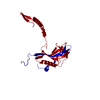
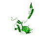
| Title | the crystal structure of recombinant baculovirus polyhedra | ||||||
 Components Components | POLYHEDRIN | ||||||
 Keywords Keywords | VIRAL PROTEIN / MICROCRYSTALS / POLYHEDRA / VIRAL OCCLUSION BODY / VIRAL CAPSID | ||||||
| Function / homology | Polyhedrin / Polyhedrin / viral occlusion body / host cell nuclear matrix / structural molecule activity / identical protein binding / Polyhedrin Function and homology information Function and homology information | ||||||
| Biological species |  AUTOGRAPHA CALIFORNICA NUCLEAR POLYHEDROSIS VIRUS AUTOGRAPHA CALIFORNICA NUCLEAR POLYHEDROSIS VIRUS | ||||||
| Method |  X-RAY DIFFRACTION / SYNCHROTRON / SIRAS / Resolution: 1.838 Å X-RAY DIFFRACTION / SYNCHROTRON / SIRAS / Resolution: 1.838 Å | ||||||
 Authors Authors | Ji, X. / Sutton, G. / Evans, G. / Axford, D. / Owen, R. / Stuart, D.I. | ||||||
 Citation Citation | Journal: Embo J. / Year: 2010 Title: How Baculovirus Polyhedra Fit Square Pegs Into Round Holes to Robustly Package Viruses. Authors: Ji, X. / Sutton, G. / Evans, G. / Axford, D. / Owen, R. / Stuart, D.I. | ||||||
| History |
|
- Structure visualization
| Structure viewer | Molecule: MolmilJmol/JSmol |
|---|
- Downloads & links
-Download
| PDBx/mmCIF format | 2wux.cif.gz | 101 KB | Display | PDBx/mmCIF format |
|---|---|---|---|---|
| PDB format | pdb2wux.ent.gz | 78.3 KB | Display | PDB format |
| PDBx/mmJSON format | 2wux.json.gz | Tree view | PDBx/mmJSON format | |
| Others |  Other downloads Other downloads |
-Validation report
| Arichive directory | https://data.pdbj.org/pub/pdb/validation_reports/wu/2wuxftp://data.pdbj.org/pub/pdb/validation_reports/wu/2wux | HTTPS FTP |
|---|
-Related structure data


-Links
 PDBj
PDBj- Assembly
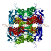
| Deposited unit | 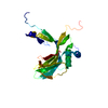
| ||||||||
|---|---|---|---|---|---|---|---|---|---|
| 1 | x 12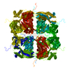
| ||||||||
| Unit cell |
|
-Components
| #1: Protein | Mass: 28736.764 Da / Num. of mol.: 1 / Mutation: YES Source method: isolated from a genetically manipulated source Source: (gene. exp.) AUTOGRAPHA CALIFORNICA NUCLEAR POLYHEDROSIS VIRUSPlasmid: PBACPAK9 / Cell line (production host): SF9 / Production host:   SPODOPTERA FRUGIPERDA (fall armyworm) / References: UniProt: P04871 SPODOPTERA FRUGIPERDA (fall armyworm) / References: UniProt: P04871 | ||
|---|---|---|---|
| #2: Water | ChemComp-HOH /  Mass: 18.015 Da / Num. of mol.: 88 / Source method: isolated from a natural source / Formula: H2O Mass: 18.015 Da / Num. of mol.: 88 / Source method: isolated from a natural source / Formula: H2O | ||
| Compound details | ENGINEERED| Has protein modification | Y | |
-Experimental details
-Experiment
| Experiment | Method: X-RAY DIFFRACTION / Number of used crystals: 17 |
|---|
- Sample preparation
| Crystal | Density Matthews: 1.57 Å3/Da / Density % sol: 21.5 % / Description: NONE |
|---|---|
| Crystal grow | pH: 7.5 / Details: pH 7.5 |
-Data collection
| Diffraction | Mean temperature: 100 K |
|---|---|
| Diffraction source | Source: SYNCHROTRON / Site: Diamond  / Beamline: I24 / Wavelength: 0.9778, 0.9778 / Beamline: I24 / Wavelength: 0.9778, 0.9778 |
| Detector | Type: RAYONIX / Detector: CCD / Date: Mar 12, 2009 / Details: MIRRORS |
| Radiation | Monochromator: DOUBLE CRYSTAL MONOCHROMATOR / Protocol: SINGLE WAVELENGTH / Monochromatic (M) / Laue (L): M / Scattering type: x-ray |
| Radiation wavelength | Wavelength: 0.9778 Å / Relative weight: 1 |
| Reflection | Resolution: 1.84→50 Å / Num. obs: 15809 / % possible obs: 100 % / Observed criterion σ(I): -1.5 / Redundancy: 14.7 % / Biso Wilson estimate: 22.75 Å2 / Rmerge(I) obs: 0.21 / Net I/σ(I): 12.1 |
| Reflection shell | Resolution: 1.84→1.91 Å / Redundancy: 10.9 % / Mean I/σ(I) obs: 1 / % possible all: 100 |
- Processing
| Software |
| |||||||||||||||||||||||||||||||||||||||||||||||||||||||||||||||||||||||||||||||||||||||||||||||||||||||||||||||||||||||||||||
|---|---|---|---|---|---|---|---|---|---|---|---|---|---|---|---|---|---|---|---|---|---|---|---|---|---|---|---|---|---|---|---|---|---|---|---|---|---|---|---|---|---|---|---|---|---|---|---|---|---|---|---|---|---|---|---|---|---|---|---|---|---|---|---|---|---|---|---|---|---|---|---|---|---|---|---|---|---|---|---|---|---|---|---|---|---|---|---|---|---|---|---|---|---|---|---|---|---|---|---|---|---|---|---|---|---|---|---|---|---|---|---|---|---|---|---|---|---|---|---|---|---|---|---|---|---|---|
| Refinement | Method to determine structure: SIRAS Starting model: NONE Resolution: 1.838→36.268 Å / SU ML: 0.22 / σ(F): 1.34 / Phase error: 20.57 / Stereochemistry target values: ML / Details: RESIDUES 3-7 WERE TRACED AS POLY ALA MODEL
| |||||||||||||||||||||||||||||||||||||||||||||||||||||||||||||||||||||||||||||||||||||||||||||||||||||||||||||||||||||||||||||
| Solvent computation | Shrinkage radii: 0.9 Å / VDW probe radii: 1.11 Å / Solvent model: FLAT BULK SOLVENT MODEL / Bsol: 94.351 Å2 / ksol: 0.445 e/Å3 | |||||||||||||||||||||||||||||||||||||||||||||||||||||||||||||||||||||||||||||||||||||||||||||||||||||||||||||||||||||||||||||
| Displacement parameters | Biso mean: 34.1 Å2 | |||||||||||||||||||||||||||||||||||||||||||||||||||||||||||||||||||||||||||||||||||||||||||||||||||||||||||||||||||||||||||||
| Refinement step | Cycle: LAST / Resolution: 1.838→36.268 Å
| |||||||||||||||||||||||||||||||||||||||||||||||||||||||||||||||||||||||||||||||||||||||||||||||||||||||||||||||||||||||||||||
| Refine LS restraints |
| |||||||||||||||||||||||||||||||||||||||||||||||||||||||||||||||||||||||||||||||||||||||||||||||||||||||||||||||||||||||||||||
| LS refinement shell |
| |||||||||||||||||||||||||||||||||||||||||||||||||||||||||||||||||||||||||||||||||||||||||||||||||||||||||||||||||||||||||||||
| Refinement TLS params. | Method: refined / Refine-ID: X-RAY DIFFRACTION
| |||||||||||||||||||||||||||||||||||||||||||||||||||||||||||||||||||||||||||||||||||||||||||||||||||||||||||||||||||||||||||||
| Refinement TLS group |
|
