National Natural Science Foundation of China (NSFC)
81520108019, 813300237
China
Chinese Academy of Sciences
2017YFC0840300
China
Citation
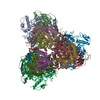
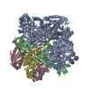
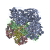
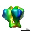
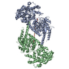
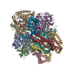
Journal: Nat Commun / Year: 2020 Title: Cryo-EM structure of trimeric Mycobacterium smegmatis succinate dehydrogenase with a membrane-anchor SdhF. Authors: Hongri Gong / Yan Gao / Xiaoting Zhou / Yu Xiao / Weiwei Wang / Yanting Tang / Shan Zhou / Yuying Zhang / Wenxin Ji / Lu Yu / Changlin Tian / Sin Man Lam / Guanghou Shui / Luke W Guddat / ...Authors: Hongri Gong / Yan Gao / Xiaoting Zhou / Yu Xiao / Weiwei Wang / Yanting Tang / Shan Zhou / Yuying Zhang / Wenxin Ji / Lu Yu / Changlin Tian / Sin Man Lam / Guanghou Shui / Luke W Guddat / Luet-Lok Wong / Quan Wang / Zihe Rao / Abstract: Diheme-containing succinate:menaquinone oxidoreductases (Sdh) are widespread in Gram-positive bacteria but little is known about the catalytic mechanisms they employ for succinate oxidation by ...Diheme-containing succinate:menaquinone oxidoreductases (Sdh) are widespread in Gram-positive bacteria but little is known about the catalytic mechanisms they employ for succinate oxidation by menaquinone. Here, we present the 2.8 Å cryo-electron microscopy structure of a Mycobacterium smegmatis Sdh, which forms a trimer. We identified the membrane-anchored SdhF as a subunit of the complex. The 3 kDa SdhF forms a single transmembrane helix and this helix plays a role in blocking the canonically proximal quinone-binding site. We also identified two distal quinone-binding sites with bound quinones. One distal binding site is formed by neighboring subunits of the complex. Our structure further reveals the electron/proton transfer pathway for succinate oxidation by menaquinone. Moreover, this study provides further structural insights into the physiological significance of a trimeric respiratory complex II. The structure of the menaquinone binding site could provide a framework for the development of Sdh-selective anti-mycobacterial drugs.
History
Deposition
Jan 29, 2020
-
Header (metadata) release
May 27, 2020
-
Map release
May 27, 2020
-
Update
Dec 24, 2025
-
Current status
Dec 24, 2025
Processing site: PDBj / Status: Released
-
Structure visualization
Movie
Surface view with section colored by density value
Number classes used: 1 / Algorithm: FOURIER SPACE / Resolution.type: BY AUTHOR / Resolution: 2.84 Å / Resolution method: FSC 0.143 CUT-OFF / Software - Name: cryoSPARC Details: cryoSPARC software was used for the reconstruction. Number images used: 461385
Initial angle assignment
Type: COMMON LINE / Software - Name: cryoSPARC
Final angle assignment
Type: COMMON LINE / Software - Name: cryoSPARC
Final 3D classification
Software - Name: cryoSPARC
+
About Yorodumi
-
News
-
Feb 9, 2022. New format data for meta-information of EMDB entries
New format data for meta-information of EMDB entries
Version 3 of the EMDB header file is now the official format.
The previous official version 1.9 will be removed from the archive.
In the structure databanks used in Yorodumi, some data are registered as the other names, "COVID-19 virus" and "2019-nCoV". Here are the details of the virus and the list of structure data.
Jan 31, 2019. EMDB accession codes are about to change! (news from PDBe EMDB page)
EMDB accession codes are about to change! (news from PDBe EMDB page)
The allocation of 4 digits for EMDB accession codes will soon come to an end. Whilst these codes will remain in use, new EMDB accession codes will include an additional digit and will expand incrementally as the available range of codes is exhausted. The current 4-digit format prefixed with “EMD-” (i.e. EMD-XXXX) will advance to a 5-digit format (i.e. EMD-XXXXX), and so on. It is currently estimated that the 4-digit codes will be depleted around Spring 2019, at which point the 5-digit format will come into force.
The EM Navigator/Yorodumi systems omit the EMD- prefix.
Related info.:Q: What is EMD? / ID/Accession-code notation in Yorodumi/EM Navigator
Yorodumi is a browser for structure data from EMDB, PDB, SASBDB, etc.
This page is also the successor to EM Navigator detail page, and also detail information page/front-end page for Omokage search.
The word "yorodu" (or yorozu) is an old Japanese word meaning "ten thousand". "mi" (miru) is to see.
Related info.:EMDB / PDB / SASBDB / Comparison of 3 databanks / Yorodumi Search / Aug 31, 2016. New EM Navigator & Yorodumi / Yorodumi Papers / Jmol/JSmol / Function and homology information / Changes in new EM Navigator and Yorodumi
 Movie
Movie Controller


 Open data
 Basic information
 Map data
 Sample
 Keywords
 Function and homology information
 Mycolicibacterium smegmatis MC2 51 (bacteria)
 Authors
 China, 3 items
  Citation

 Structure visualization
 Downloads & links
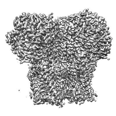 emd_0981.png
 http://ftp.pdbj.org/pub/emdb/structures/EMD-0981


 Z (Sec.)
 Y (Row.)
 X (Col.)


 Sample components


 Processing
 Electron microscopy
 FIELD EMISSION GUN
