| Entry | Database: PDB / ID: 5sul
|
|---|
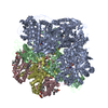
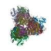
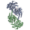
| Title | Inhibited state structure of yGsy2p |
|---|
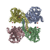
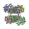
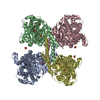
 Components Components | Glycogen [starch] synthase isoform 2 |
|---|
 Keywords Keywords | TRANSFERASE / Glycogen synthase Inhibited state / Phosphorylation |
|---|
| Function / homology |  Function and homology information Function and homology information
Glycogen synthesis / glycogen binding / glycogen(starch) synthase / alpha-1,4-glucan glucosyltransferase (UDP-glucose donor) activity / glycogen granule / glycogen biosynthetic process / identical protein binding / nucleus / cytoplasm / cytosolSimilarity search - Function |
|---|
| Biological species |   Saccharomyces cerevisiae (brewer's yeast) Saccharomyces cerevisiae (brewer's yeast) |
|---|
| Method |  X-RAY DIFFRACTION / SYNCHROTRON / MOLECULAR REPLACEMENT / Resolution: 3.3 Å X-RAY DIFFRACTION / SYNCHROTRON / MOLECULAR REPLACEMENT / Resolution: 3.3 Å |
|---|
 Authors Authors | Mahalingan, K.K. / Hurley, T.D. |
|---|
| Funding support |  United States, 2items United States, 2items | Organization | Grant number | Country |
|---|
| National Institutes of Health/National Institute of Diabetes and Digestive and Kidney Disease (NIH/NIDDK) | | United States | | National Institutes of Health/National Institute of Neurological Disorders and Stroke (NIH/NINDS) | | United States |
|
|---|
 Citation Citation | Journal: Biochemistry / Year: 2017
Title: Redox Switch for the Inhibited State of Yeast Glycogen Synthase Mimics Regulation by Phosphorylation.
Authors: Mahalingan, K.K. / Baskaran, S. / DePaoli-Roach, A.A. / Roach, P.J. / Hurley, T.D. |
|---|
| History | | Deposition | Aug 3, 2016 | Deposition site: RCSB / Processing site: RCSB |
|---|
| Revision 1.0 | Jun 14, 2017 | Provider: repository / Type: Initial release |
|---|
| Revision 1.1 | Sep 6, 2017 | Group: Author supporting evidence / Category: pdbx_audit_support |
|---|
| Revision 1.2 | Dec 18, 2019 | Group: Author supporting evidence / Category: pdbx_audit_support / Item: _pdbx_audit_support.funding_organization |
|---|
| Revision 1.3 | Oct 4, 2023 | Group: Data collection / Database references / Refinement description
Category: chem_comp_atom / chem_comp_bond ...chem_comp_atom / chem_comp_bond / database_2 / pdbx_initial_refinement_model / struct_ncs_dom_lim
Item: _database_2.pdbx_DOI / _database_2.pdbx_database_accession ..._database_2.pdbx_DOI / _database_2.pdbx_database_accession / _struct_ncs_dom_lim.beg_auth_comp_id / _struct_ncs_dom_lim.beg_label_asym_id / _struct_ncs_dom_lim.beg_label_comp_id / _struct_ncs_dom_lim.beg_label_seq_id / _struct_ncs_dom_lim.end_auth_comp_id / _struct_ncs_dom_lim.end_label_asym_id / _struct_ncs_dom_lim.end_label_comp_id / _struct_ncs_dom_lim.end_label_seq_id |
|---|
|
|---|
 Movie
Movie Controller


 Open data
 Basic information
 Structure visualization
 Downloads & links
 Other downloads
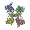


 PDBj
PDBj


 Assembly
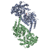


 Type: RNA linking / Mass: 404.161 Da / Num. of mol.: 1 / Source method: obtained synthetically / Formula: C9H14N2O12P2 / Comment: UDP*YM
 Mass: 324.181 Da / Num. of mol.: 1 / Source method: obtained synthetically / Formula: C9H13N2O9P
 Sample preparation
 Processing