 Movie
Movie Controller


+ Open data

- Basic information
| Entry | Database: PDB / ID: 6lum | ||||||||||||||||||||||||||||||||||||||||||||||||||||||||||||||||||||||||||||||||||||||||||
|---|---|---|---|---|---|---|---|---|---|---|---|---|---|---|---|---|---|---|---|---|---|---|---|---|---|---|---|---|---|---|---|---|---|---|---|---|---|---|---|---|---|---|---|---|---|---|---|---|---|---|---|---|---|---|---|---|---|---|---|---|---|---|---|---|---|---|---|---|---|---|---|---|---|---|---|---|---|---|---|---|---|---|---|---|---|---|---|---|---|---|---|
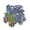
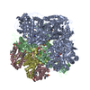
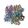
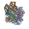
| Title | Structure of Mycobacterium smegmatis succinate dehydrogenase 2 | ||||||||||||||||||||||||||||||||||||||||||||||||||||||||||||||||||||||||||||||||||||||||||
 Components Components | (Succinate dehydrogenase subunit ...) x 5 | ||||||||||||||||||||||||||||||||||||||||||||||||||||||||||||||||||||||||||||||||||||||||||
 Keywords Keywords | OXIDOREDUCTASE / electron transfer chain / trimer | ||||||||||||||||||||||||||||||||||||||||||||||||||||||||||||||||||||||||||||||||||||||||||
| Function / homology |  Function and homology information Function and homology information: / steroid dehydrogenase activity, acting on the CH-CH group of donors / succinate dehydrogenase (quinone) activity / succinate dehydrogenase / 3 iron, 4 sulfur cluster binding / steroid metabolic process / tricarboxylic acid cycle / electron transport chain / 2 iron, 2 sulfur cluster binding / flavin adenine dinucleotide binding ...: / steroid dehydrogenase activity, acting on the CH-CH group of donors / succinate dehydrogenase (quinone) activity / succinate dehydrogenase / 3 iron, 4 sulfur cluster binding / steroid metabolic process / tricarboxylic acid cycle / electron transport chain / 2 iron, 2 sulfur cluster binding / flavin adenine dinucleotide binding / 4 iron, 4 sulfur cluster binding / electron transfer activity / oxidoreductase activity / membrane / metal ion binding Similarity search - Function | ||||||||||||||||||||||||||||||||||||||||||||||||||||||||||||||||||||||||||||||||||||||||||
| Biological species |  Mycolicibacterium smegmatis MC2 51 (bacteria) Mycolicibacterium smegmatis MC2 51 (bacteria) | ||||||||||||||||||||||||||||||||||||||||||||||||||||||||||||||||||||||||||||||||||||||||||
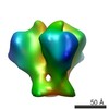
| Method | ELECTRON MICROSCOPY / single particle reconstruction / cryo EM / Resolution: 2.84 Å | ||||||||||||||||||||||||||||||||||||||||||||||||||||||||||||||||||||||||||||||||||||||||||
 Authors Authors | Gao, Y. / Gong, H. / Zhou, X. / Xiao, Y. / Wang, W. / Ji, W. / Wang, Q. / Rao, Z. | ||||||||||||||||||||||||||||||||||||||||||||||||||||||||||||||||||||||||||||||||||||||||||
| Funding support |  China, 3items China, 3items
| ||||||||||||||||||||||||||||||||||||||||||||||||||||||||||||||||||||||||||||||||||||||||||
 Citation Citation | Journal: Nat Commun / Year: 2020 Title: Cryo-EM structure of trimeric Mycobacterium smegmatis succinate dehydrogenase with a membrane-anchor SdhF. Authors: Hongri Gong / Yan Gao / Xiaoting Zhou / Yu Xiao / Weiwei Wang / Yanting Tang / Shan Zhou / Yuying Zhang / Wenxin Ji / Lu Yu / Changlin Tian / Sin Man Lam / Guanghou Shui / Luke W Guddat / ...Authors: Hongri Gong / Yan Gao / Xiaoting Zhou / Yu Xiao / Weiwei Wang / Yanting Tang / Shan Zhou / Yuying Zhang / Wenxin Ji / Lu Yu / Changlin Tian / Sin Man Lam / Guanghou Shui / Luke W Guddat / Luet-Lok Wong / Quan Wang / Zihe Rao /   Abstract: Diheme-containing succinate:menaquinone oxidoreductases (Sdh) are widespread in Gram-positive bacteria but little is known about the catalytic mechanisms they employ for succinate oxidation by ...Diheme-containing succinate:menaquinone oxidoreductases (Sdh) are widespread in Gram-positive bacteria but little is known about the catalytic mechanisms they employ for succinate oxidation by menaquinone. Here, we present the 2.8 Å cryo-electron microscopy structure of a Mycobacterium smegmatis Sdh, which forms a trimer. We identified the membrane-anchored SdhF as a subunit of the complex. The 3 kDa SdhF forms a single transmembrane helix and this helix plays a role in blocking the canonically proximal quinone-binding site. We also identified two distal quinone-binding sites with bound quinones. One distal binding site is formed by neighboring subunits of the complex. Our structure further reveals the electron/proton transfer pathway for succinate oxidation by menaquinone. Moreover, this study provides further structural insights into the physiological significance of a trimeric respiratory complex II. The structure of the menaquinone binding site could provide a framework for the development of Sdh-selective anti-mycobacterial drugs. | ||||||||||||||||||||||||||||||||||||||||||||||||||||||||||||||||||||||||||||||||||||||||||
| History |
|
- Structure visualization
| Movie |
Movie viewer |
|---|---|
| Structure viewer | Molecule: MolmilJmol/JSmol |
- Downloads & links
-Download
| PDBx/mmCIF format | 6lum.cif.gz | 597.8 KB | Display | PDBx/mmCIF format |
|---|---|---|---|---|
| PDB format | pdb6lum.ent.gz | 480.9 KB | Display | PDB format |
| PDBx/mmJSON format | 6lum.json.gz | Tree view | PDBx/mmJSON format | |
| Others |  Other downloads Other downloads |
-Validation report
| Arichive directory | https://data.pdbj.org/pub/pdb/validation_reports/lu/6lumftp://data.pdbj.org/pub/pdb/validation_reports/lu/6lum | HTTPS FTP |
|---|
-Related structure data
| Related structure data |  0981MC M: map data used to model this data C: citing same article ( |
|---|---|
| Similar structure data |


-Links
 PDBj
PDBj


- Assembly
| Deposited unit | 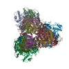
|
|---|---|
| 1 |
|
-Components
-Succinate dehydrogenase subunit ... , 5 types, 15 molecules CGMDHNEIOAJPBKQ
| #1: Protein | Mass: 15644.627 Da / Num. of mol.: 3 / Source method: isolated from a natural source Source: (natural) Mycolicibacterium smegmatis MC2 51 (bacteria)Strain: MC2 51 / References: UniProt: A0QT10*PLUS #2: Protein | Mass: 19280.400 Da / Num. of mol.: 3 / Source method: isolated from a natural source Source: (natural) Mycolicibacterium smegmatis MC2 51 (bacteria)Strain: MC2 51 / References: UniProt: A0QT09*PLUS #3: Protein/peptide | Mass: 3707.443 Da / Num. of mol.: 3 / Source method: isolated from a natural source Source: (natural) Mycolicibacterium smegmatis MC2 51 (bacteria)Strain: MC2 51 / References: UniProt: A0R4D1*PLUS #4: Protein | Mass: 64476.754 Da / Num. of mol.: 3 / Source method: isolated from a natural source Source: (natural) Mycolicibacterium smegmatis MC2 51 (bacteria)Strain: MC2 51 / References: UniProt: A0QT08*PLUS #5: Protein | Mass: 29245.760 Da / Num. of mol.: 3 / Source method: isolated from a natural source Source: (natural) Mycolicibacterium smegmatis MC2 51 (bacteria)Strain: MC2 51 / References: UniProt: A0QT07*PLUS |
|---|
-Non-polymers , 10 types, 39 molecules 


| #6: Chemical | ChemComp-PEV / (  Mass: 720.012 Da / Num. of mol.: 6 / Source method: obtained synthetically / Formula: C39H78NO8P / Feature type: SUBJECT OF INVESTIGATION / Comment: POPE, phospholipid*YM Mass: 720.012 Da / Num. of mol.: 6 / Source method: obtained synthetically / Formula: C39H78NO8P / Feature type: SUBJECT OF INVESTIGATION / Comment: POPE, phospholipid*YM#7: Chemical | ChemComp-HEM /  Mass: 616.487 Da / Num. of mol.: 6 / Source method: obtained synthetically / Formula: C34H32FeN4O4 / Feature type: SUBJECT OF INVESTIGATION Mass: 616.487 Da / Num. of mol.: 6 / Source method: obtained synthetically / Formula: C34H32FeN4O4 / Feature type: SUBJECT OF INVESTIGATION#8: Chemical | ChemComp-MQ9 /  Mass: 785.233 Da / Num. of mol.: 6 / Source method: obtained synthetically / Formula: C56H80O2 / Feature type: SUBJECT OF INVESTIGATION Mass: 785.233 Da / Num. of mol.: 6 / Source method: obtained synthetically / Formula: C56H80O2 / Feature type: SUBJECT OF INVESTIGATION#9: Chemical |  Mass: 1464.043 Da / Num. of mol.: 3 / Source method: obtained synthetically / Formula: C81H156O17P2 / Feature type: SUBJECT OF INVESTIGATION / Comment: phospholipid*YM Mass: 1464.043 Da / Num. of mol.: 3 / Source method: obtained synthetically / Formula: C81H156O17P2 / Feature type: SUBJECT OF INVESTIGATION / Comment: phospholipid*YM#10: Chemical |  Mass: 648.891 Da / Num. of mol.: 3 / Source method: obtained synthetically / Formula: C35H69O8P / Comment: phospholipid*YM Mass: 648.891 Da / Num. of mol.: 3 / Source method: obtained synthetically / Formula: C35H69O8P / Comment: phospholipid*YM#11: Chemical |  Mass: 836.061 Da / Num. of mol.: 3 / Source method: obtained synthetically / Formula: C43H80O13P / Feature type: SUBJECT OF INVESTIGATION Mass: 836.061 Da / Num. of mol.: 3 / Source method: obtained synthetically / Formula: C43H80O13P / Feature type: SUBJECT OF INVESTIGATION#12: Chemical |  Mass: 785.550 Da / Num. of mol.: 3 / Source method: obtained synthetically / Formula: C27H33N9O15P2 / Feature type: SUBJECT OF INVESTIGATION / Comment: FAD*YM Mass: 785.550 Da / Num. of mol.: 3 / Source method: obtained synthetically / Formula: C27H33N9O15P2 / Feature type: SUBJECT OF INVESTIGATION / Comment: FAD*YM#13: Chemical |  Mass: 175.820 Da / Num. of mol.: 3 / Source method: obtained synthetically / Formula: Fe2S2 Mass: 175.820 Da / Num. of mol.: 3 / Source method: obtained synthetically / Formula: Fe2S2#14: Chemical |  Mass: 351.640 Da / Num. of mol.: 3 / Source method: obtained synthetically / Formula: Fe4S4 Mass: 351.640 Da / Num. of mol.: 3 / Source method: obtained synthetically / Formula: Fe4S4#15: Chemical |  Mass: 295.795 Da / Num. of mol.: 3 / Source method: obtained synthetically / Formula: Fe3S4 Mass: 295.795 Da / Num. of mol.: 3 / Source method: obtained synthetically / Formula: Fe3S4 |
|---|
-Details
| Has ligand of interest | Y |
|---|---|
| Has protein modification | N |
-Experimental details
-Experiment
| Experiment | Method: ELECTRON MICROSCOPY |
|---|---|
| EM experiment | Aggregation state: PARTICLE / 3D reconstruction method: single particle reconstruction |
- Sample preparation
| Component | Name: Mycobaterium smegmatis Succinate dehydrogenase 2 / Type: COMPLEX / Entity ID: #1-#5 / Source: NATURAL | ||||||||||||||||||||
|---|---|---|---|---|---|---|---|---|---|---|---|---|---|---|---|---|---|---|---|---|---|
| Molecular weight | Value: 0.4 MDa / Experimental value: YES | ||||||||||||||||||||
| Source (natural) | Organism: Mycolicibacterium smegmatis MC2 51 (bacteria) / Strain: mc2 51 | ||||||||||||||||||||
| Buffer solution | pH: 7.4 / Details: Solutions were made fresh | ||||||||||||||||||||
| Buffer component |
| ||||||||||||||||||||
| Specimen | Conc.: 5 mg/ml / Embedding applied: NO / Shadowing applied: NO / Staining applied: NO / Vitrification applied: YES / Details: This sample was mono disperse. | ||||||||||||||||||||
| Specimen support | Grid material: COPPER / Grid mesh size: 300 divisions/in. / Grid type: Quantifoil R1.2/1.3 | ||||||||||||||||||||
| Vitrification | Instrument: FEI VITROBOT MARK II / Cryogen name: ETHANE / Humidity: 100 % / Chamber temperature: 281 K |
- Electron microscopy imaging
| Experimental equipment |  Model: Titan Krios / Image courtesy: FEI Company |
|---|---|
| Microscopy | Model: FEI TITAN KRIOS |
| Electron gun | Electron source:  FIELD EMISSION GUN / Accelerating voltage: 300 kV / Illumination mode: SPOT SCAN FIELD EMISSION GUN / Accelerating voltage: 300 kV / Illumination mode: SPOT SCAN |
| Electron lens | Mode: BRIGHT FIELD / Cs: 2.7 mm / C2 aperture diameter: 70 µm |
| Image recording | Average exposure time: 2 sec. / Electron dose: 50 e/Å2 / Film or detector model: GATAN K3 (6k x 4k) |
- Processing
| Software | Name: PHENIX / Version: 1.16_3549: / Classification: refinement | ||||||||||||||||||||||||||||
|---|---|---|---|---|---|---|---|---|---|---|---|---|---|---|---|---|---|---|---|---|---|---|---|---|---|---|---|---|---|
| EM software |
| ||||||||||||||||||||||||||||
| CTF correction | Type: PHASE FLIPPING AND AMPLITUDE CORRECTION | ||||||||||||||||||||||||||||
| 3D reconstruction | Resolution: 2.84 Å / Resolution method: FSC 0.143 CUT-OFF / Num. of particles: 461385 / Algorithm: FOURIER SPACE Details: cryoSPARC software was used for the reconstruction. Num. of class averages: 1 / Symmetry type: POINT | ||||||||||||||||||||||||||||
| Refinement | Highest resolution: 2.84 Å | ||||||||||||||||||||||||||||
| Refine LS restraints |
|
