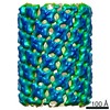
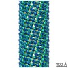
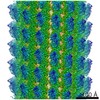
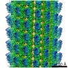
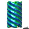
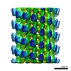
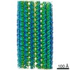
Journal: J Am Chem Soc / Year: 2019 Title: Chemically Controlled Helical Polymorphism in Protein Tubes by Selective Modulation of Supramolecular Interactions. Authors: Zhen Li / Shuyu Chen / Chendi Gao / Zhiwei Yang / Kuo-Chih Shih / Zdravko Kochovski / Guang Yang / Lu Gou / Mu-Ping Nieh / Ming Jiang / Lei Zhang / Guosong Chen / Abstract: Polymorphism has been the subject of investigation across different research disciplines. In biology, polymorphism could be interpreted in such a way that discrete biomacromolecules can adopt ...Polymorphism has been the subject of investigation across different research disciplines. In biology, polymorphism could be interpreted in such a way that discrete biomacromolecules can adopt diversiform specific conformations/packing arrangement, and this polymorph-dependent property is essential for many biochemical processes. For example, bacterial flagellar filament, composed of flagellin, switches between different supercoiled state allowing the bacteria to swim and tumble. However, in artificial supramolecular systems, it is often challenging to achieve polymorph control and prediction, and in most cases, two or more concomitant polymorphs of similar formation energies coexist. Here, we show that a tetrameric protein with properly oriented binding sites on its surface can arrange into diverse protein tubes with distinct helical parameters by adding specifically designed inducing ligands. We examined several parameters of the ligand that would influence the protein tube formation and found that the flexibility of the ligand linker and the dimerization pose of the ligand complex is critical for the successful production of the tubes and eventually influence the specific helical polymorphs of the formed tubes. A surface lattice accommodation model was further developed to rationalize the geometrical relationship between each helical tube type. Molecular simulation was used to elucidate the interactions between ligands and SBA and molecular basis for polymorphic switching of the protein tubes. Moreover, the kinetics of structural formation was studied and the ligand design was found that can affect the kinetics of the protein polymerization pathway. In short, our designed protein tubes serves as an enlightening system for understanding how a protein polymer composed of a single protein switches among different helical states.
History
Deposition
Aug 10, 2019
-
Header (metadata) release
Jun 24, 2020
-
Map release
Jun 24, 2020
-
Update
Jun 24, 2020
-
Current status
Jun 24, 2020
Processing site: PDBj / Status: Released
-
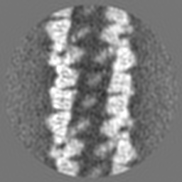
Structure visualization
Movie
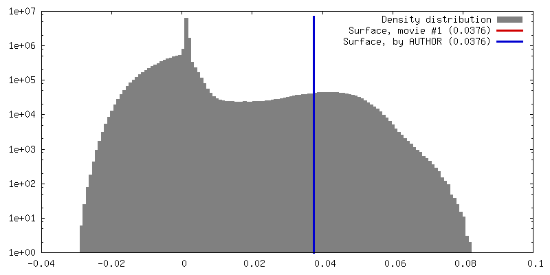
Surface view with section colored by density value
In the structure databanks used in Yorodumi, some data are registered as the other names, "COVID-19 virus" and "2019-nCoV". Here are the details of the virus and the list of structure data.
Jan 31, 2019. EMDB accession codes are about to change! (news from PDBe EMDB page)
EMDB accession codes are about to change! (news from PDBe EMDB page)
The allocation of 4 digits for EMDB accession codes will soon come to an end. Whilst these codes will remain in use, new EMDB accession codes will include an additional digit and will expand incrementally as the available range of codes is exhausted. The current 4-digit format prefixed with “EMD-” (i.e. EMD-XXXX) will advance to a 5-digit format (i.e. EMD-XXXXX), and so on. It is currently estimated that the 4-digit codes will be depleted around Spring 2019, at which point the 5-digit format will come into force.
The EM Navigator/Yorodumi systems omit the EMD- prefix.
Related info.:Q: What is EMD? / ID/Accession-code notation in Yorodumi/EM Navigator
Yorodumi is a browser for structure data from EMDB, PDB, SASBDB, etc.
This page is also the successor to EM Navigator detail page, and also detail information page/front-end page for Omokage search.
The word "yorodu" (or yorozu) is an old Japanese word meaning "ten thousand". "mi" (miru) is to see.
Related info.:EMDB / PDB / SASBDB / Comparison of 3 databanks / Yorodumi Search / Aug 31, 2016. New EM Navigator & Yorodumi / Yorodumi Papers / Jmol/JSmol / Function and homology information / Changes in new EM Navigator and Yorodumi
 Movie
Movie Controller

 Yorodumi
 Open data
 Basic information
 Map data
 Sample
 Authors
 China, 1 items
  Citation

 Structure visualization
 Movie viewer
 Downloads & links
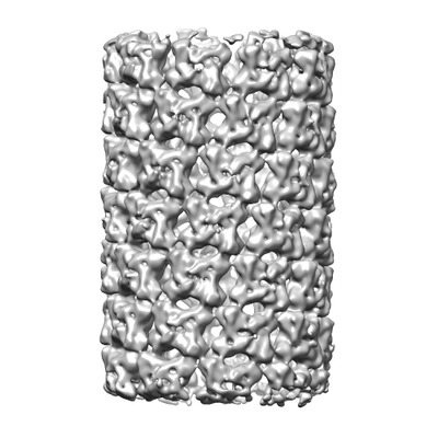 emd_0736.png
 http://ftp.pdbj.org/pub/emdb/structures/EMD-0736
 Z (Sec.)
 Y (Row.)
 X (Col.)


 Sample components
 Processing
 Electron microscopy
 FIELD EMISSION GUN

