 Movie
Movie Controller

[English] 日本語
 Yorodumi
- EMDB-0399: Electron microscopy snapshots of single particles from single cells -
+ Open data

- Basic information
| Entry | Database: EMDB / ID: EMD-0399 | |||||||||
|---|---|---|---|---|---|---|---|---|---|---|
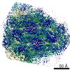
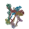
| Title | Electron microscopy snapshots of single particles from single cells | |||||||||
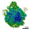
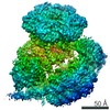
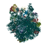
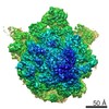
 Map data Map data | Reconstruction of 60S ribosome | |||||||||
 Sample Sample |
| |||||||||
| Biological species |  | |||||||||
| Method | single particle reconstruction / negative staining / Resolution: 34.0 Å | |||||||||
 Authors Authors | Yi X / Verbeke EJ / Yiran C / Dickinson DJ / Taylor DW | |||||||||
 Citation Citation | Journal: J Biol Chem / Year: 2019 Title: Electron microscopy snapshots of single particles from single cells. Authors: Xiunan Yi / Eric J Verbeke / Yiran Chang / Daniel J Dickinson / David W Taylor /  Abstract: Cryo-electron microscopy (cryo-EM) has become an indispensable tool for structural studies of biological macromolecules. Two additional predominant methods are available for studying the ...Cryo-electron microscopy (cryo-EM) has become an indispensable tool for structural studies of biological macromolecules. Two additional predominant methods are available for studying the architectures of multiprotein complexes: 1) single-particle analysis of purified samples and 2) tomography of whole cells or cell sections. The former can produce high-resolution structures but is limited to highly purified samples, whereas the latter can capture proteins in their native state but has a low signal-to-noise ratio and yields lower-resolution structures. Here, we present a simple, adaptable method combining microfluidic single-cell extraction with single-particle analysis by EM to characterize protein complexes from individual embryos. Using this approach, we uncover 3D structures of ribosomes directly from single embryo extracts. Moreover, we investigated structural dynamics during development by counting the number of ribosomes per polysome in early and late embryos. This approach has significant potential applications for counting protein complexes and studying protein architectures from single cells in developmental, evolutionary, and disease contexts. | |||||||||
| History |
|
- Structure visualization
| Movie |
 Movie viewer Movie viewer |
|---|---|
| Structure viewer | EM map: SurfViewMolmilJmol/JSmol |
| Supplemental images |

- Downloads & links
-EMDB archive
| Map data | emd_0399.map.gz | 1.4 MB | EMDB map data format | |
|---|---|---|---|---|
| Header (meta data) | emd-0399-v30.xmlemd-0399.xml | 8.2 KB 8.2 KB | Display Display | EMDB header |
| Images | 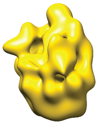 emd_0399.png emd_0399.png | 98.6 KB | ||
| Archive directory |  http://ftp.pdbj.org/pub/emdb/structures/EMD-0399ftp://ftp.pdbj.org/pub/emdb/structures/EMD-0399 http://ftp.pdbj.org/pub/emdb/structures/EMD-0399ftp://ftp.pdbj.org/pub/emdb/structures/EMD-0399 | HTTPS FTP |
-Validation report
| Summary document | emd_0399_validation.pdf.gz | 78.8 KB | Display | EMDB validaton report |
|---|---|---|---|---|
| Full document | emd_0399_full_validation.pdf.gz | 77.9 KB | Display | |
| Data in XML | emd_0399_validation.xml.gz | 495 B | Display | |
| Arichive directory | https://ftp.pdbj.org/pub/emdb/validation_reports/EMD-0399ftp://ftp.pdbj.org/pub/emdb/validation_reports/EMD-0399 | HTTPS FTP |
-Related structure data

-Links
| EMDB pages | EMDB (EBI/PDBe) / EMDataResource |
|---|---|
| Related items in Molecule of the Month |

-Map
| File | Download / File: emd_0399.map.gz / Format: CCP4 / Size: 6.6 MB / Type: IMAGE STORED AS FLOATING POINT NUMBER (4 BYTES) | ||||||||||||||||||||||||||||||||||||||||||||||||||||||||||||
|---|---|---|---|---|---|---|---|---|---|---|---|---|---|---|---|---|---|---|---|---|---|---|---|---|---|---|---|---|---|---|---|---|---|---|---|---|---|---|---|---|---|---|---|---|---|---|---|---|---|---|---|---|---|---|---|---|---|---|---|---|---|
| Annotation | Reconstruction of 60S ribosome | ||||||||||||||||||||||||||||||||||||||||||||||||||||||||||||
| Projections & slices | Image control
Images are generated by Spider. | ||||||||||||||||||||||||||||||||||||||||||||||||||||||||||||
| Voxel size | X=Y=Z: 3.6 Å | ||||||||||||||||||||||||||||||||||||||||||||||||||||||||||||
| Density |
| ||||||||||||||||||||||||||||||||||||||||||||||||||||||||||||
| Symmetry | Space group: 1 | ||||||||||||||||||||||||||||||||||||||||||||||||||||||||||||
| Details | EMDB XML:
CCP4 map header:
| ||||||||||||||||||||||||||||||||||||||||||||||||||||||||||||
 Z (Sec.)
 Y (Row.)
 X (Col.)


-Supplemental data
- Sample components
-Entire : 60S ribosome from single cells
| Entire | Name: 60S ribosome from single cells |
|---|---|
| Components |
|
-Supramolecule #1: 60S ribosome from single cells
| Supramolecule | Name: 60S ribosome from single cells / type: complex / ID: 1 / Parent: 0 |
|---|---|
| Source (natural) | Organism: |
| Molecular weight | Theoretical: 1 MDa |
-Experimental details
-Structure determination
| Method | negative staining |
|---|---|
 Processing Processing | single particle reconstruction |
| Aggregation state | particle |
-Sample preparation
| Concentration | 0.02 mg/mL |
|---|---|
| Buffer | pH: 7.4 |
| Staining | Type: NEGATIVE / Material: Uranyl Acetate |
| Grid | Details: unspecified |
- Electron microscopy
| Microscope | JEOL 2010F |
|---|---|
| Image recording | Film or detector model: GATAN MULTISCAN / Average electron dose: 30.0 e/Å2 |
| Electron beam | Acceleration voltage: 200 kV / Electron source:  FIELD EMISSION GUN FIELD EMISSION GUN |
| Electron optics | Illumination mode: FLOOD BEAM / Imaging mode: BRIGHT FIELD |
-Image processing
| Particle selection | Number selected: 30000 |
|---|---|
| Final reconstruction | Applied symmetry - Point group: C1 (asymmetric) / Resolution.type: BY AUTHOR / Resolution: 34.0 Å / Resolution method: FSC 0.5 CUT-OFF / Number images used: 3400 |
| Initial angle assignment | Type: PROJECTION MATCHING |
| Final angle assignment | Type: PROJECTION MATCHING |
