 Movie
Movie Controller

[English] 日本語
 Yorodumi
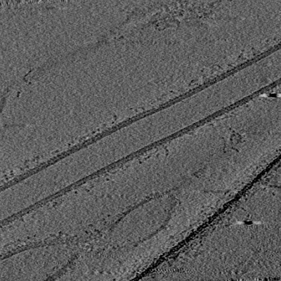
- EMDB-0307: Representative tomogram of the COPII in vitro reconstitution used... -
+ Open data

- Basic information
| Entry | Database: EMDB / ID: EMD-0307 | |||||||||
|---|---|---|---|---|---|---|---|---|---|---|
| Title | Representative tomogram of the COPII in vitro reconstitution used for subtomogram averaging | |||||||||
 Map data Map data | *** This is a dummy file *** Representative unbinned 3D-CTF corrected tomogram used for subtomogram averaging of COPII assembled on membranes. | |||||||||
 Sample Sample |
| |||||||||
| Biological species |  | |||||||||
| Method | electron tomography / cryo EM | |||||||||
 Authors Authors | Zanetti G / Hutchings J / Hagen WJH | |||||||||
| Funding support |  United Kingdom, 2 items United Kingdom, 2 items
| |||||||||
 Citation Citation | Journal: Nat Commun / Year: 2018 Title: Subtomogram averaging of COPII assemblies reveals how coat organization dictates membrane shape. Authors: Joshua Hutchings / Viktoriya Stancheva / Elizabeth A Miller / Giulia Zanetti / Abstract: Eukaryotic cells employ membrane-bound carriers to transport cargo between compartments in a process essential to cell functionality. Carriers are generated by coat complexes that couple cargo ...Eukaryotic cells employ membrane-bound carriers to transport cargo between compartments in a process essential to cell functionality. Carriers are generated by coat complexes that couple cargo capture to membrane deformation. The COPII coat mediates export from the endoplasmic reticulum by assembling in inner and outer layers, yielding carriers of variable shape and size that allow secretion of thousands of diverse cargo. Despite detailed understanding of COPII subunits, the molecular mechanisms of coat assembly and membrane deformation are unclear. Here we present a 4.9 Å cryo-tomography subtomogram averaging structure of in vitro-reconstituted membrane-bound inner coat. We show that the outer coat (Sec13-Sec31) bridges inner coat subunits (Sar1-Sec23-Sec24), promoting their assembly into a tight lattice. We directly visualize the membrane-embedded Sar1 amphipathic helix, revealing that lattice formation induces parallel helix insertions, yielding tubular curvature. We propose that regulators like the procollagen receptor TANGO1 modulate this mechanism to determine vesicle shape and size. | |||||||||
| History |
|
- Structure visualization
| Movie |
 Movie viewer Movie viewer |
|---|---|
| Supplemental images |
- Downloads & links
-EMDB archive
| Map data | emd_0307.map.gz | 40.8 GB | EMDB map data format | |
|---|---|---|---|---|
| Header (meta data) | emd-0307-v30.xmlemd-0307.xml | 8.9 KB 8.9 KB | Display Display | EMDB header |
| Images |  emd_0307.png emd_0307.png | 125.1 KB | ||
| Archive directory |  http://ftp.pdbj.org/pub/emdb/structures/EMD-0307ftp://ftp.pdbj.org/pub/emdb/structures/EMD-0307 http://ftp.pdbj.org/pub/emdb/structures/EMD-0307ftp://ftp.pdbj.org/pub/emdb/structures/EMD-0307 | HTTPS FTP |
-Validation report
| Summary document | emd_0307_validation.pdf.gz | 136.1 KB | Display | EMDB validaton report |
|---|---|---|---|---|
| Full document | emd_0307_full_validation.pdf.gz | 135.2 KB | Display | |
| Data in XML | emd_0307_validation.xml.gz | 3.4 KB | Display | |
| Arichive directory | https://ftp.pdbj.org/pub/emdb/validation_reports/EMD-0307ftp://ftp.pdbj.org/pub/emdb/validation_reports/EMD-0307 | HTTPS FTP |
-Related structure data

-Links
| EMDB pages | EMDB (EBI/PDBe) / EMDataResource |
|---|
-Map
| File | Download / File: emd_0307.map.gz / Format: CCP4 / Size: 4.9 KB / Type: IMAGE STORED AS SIGNED INTEGER (2 BYTES) | ||||||||||||||||||||||||||||||||||||||||||||||||||||||||||||
|---|---|---|---|---|---|---|---|---|---|---|---|---|---|---|---|---|---|---|---|---|---|---|---|---|---|---|---|---|---|---|---|---|---|---|---|---|---|---|---|---|---|---|---|---|---|---|---|---|---|---|---|---|---|---|---|---|---|---|---|---|---|
| Annotation | *** This is a dummy file *** Representative unbinned 3D-CTF corrected tomogram used for subtomogram averaging of COPII assembled on membranes. | ||||||||||||||||||||||||||||||||||||||||||||||||||||||||||||
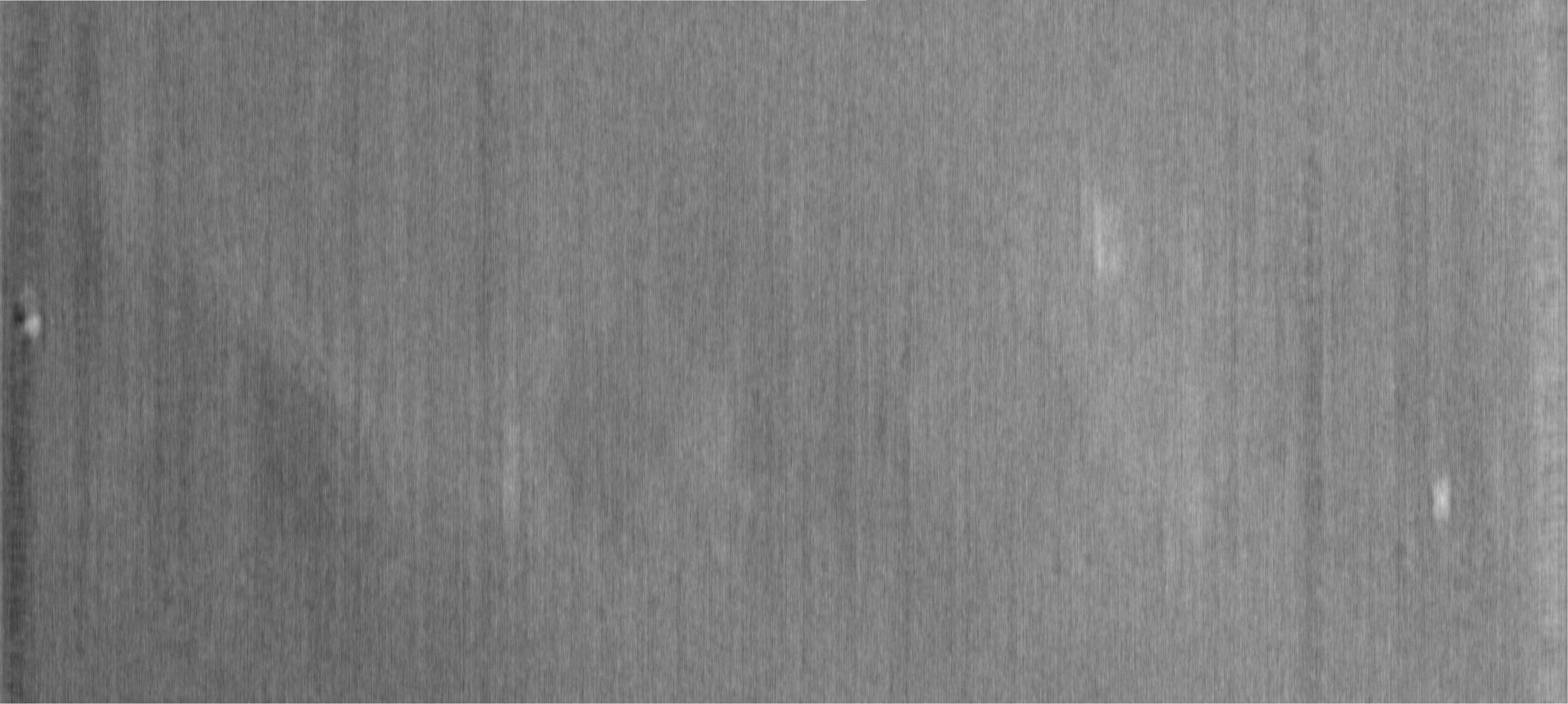
| Projections & slices | Image control
Images are generated by Spider. generated in cubic-lattice coordinate | ||||||||||||||||||||||||||||||||||||||||||||||||||||||||||||
| Voxel size |
| ||||||||||||||||||||||||||||||||||||||||||||||||||||||||||||
| Density |
| ||||||||||||||||||||||||||||||||||||||||||||||||||||||||||||
| Symmetry | Space group: 0 | ||||||||||||||||||||||||||||||||||||||||||||||||||||||||||||
| Details | EMDB XML:
CCP4 map header:
| ||||||||||||||||||||||||||||||||||||||||||||||||||||||||||||
 Z (Sec.)
 Y (Row.)
 X (Col.)


-Supplemental data
- Sample components
-Entire : In vitro reconstitution of COPII membrane deformation with giant ...
| Entire | Name: In vitro reconstitution of COPII membrane deformation with giant unilamellar vesicles (GUVs). |
|---|---|
| Components |
|
-Supramolecule #1: In vitro reconstitution of COPII membrane deformation with giant ...
| Supramolecule | Name: In vitro reconstitution of COPII membrane deformation with giant unilamellar vesicles (GUVs). type: complex / ID: 1 / Parent: 0 |
|---|---|
| Source (natural) | Organism: |
| Recombinant expression | Organism:   Spodoptera frugiperda (fall armyworm) Spodoptera frugiperda (fall armyworm) |
-Experimental details
-Structure determination
| Method | cryo EM |
|---|---|
 Processing Processing | electron tomography |
| Aggregation state | helical array |
-Sample preparation
| Buffer | pH: 6.8 |
|---|---|
| Vitrification | Cryogen name: ETHANE / Chamber humidity: 100 % / Chamber temperature: 277 K / Instrument: FEI VITROBOT MARK II |
| Sectioning | Other: NO SECTIONING |
| Fiducial marker | Manufacturer: BBI / Diameter: 5 nm |
- Electron microscopy
| Microscope | FEI TITAN KRIOS |
|---|---|
| Image recording | Film or detector model: GATAN K2 SUMMIT (4k x 4k) / Detector mode: COUNTING / Average electron dose: 3.6 e/Å2 |
| Electron beam | Acceleration voltage: 300 kV / Electron source:  FIELD EMISSION GUN FIELD EMISSION GUN |
| Electron optics | C2 aperture diameter: 50.0 µm / Illumination mode: OTHER / Imaging mode: BRIGHT FIELD / Cs: 2.7 mm / Nominal defocus max: 0.0035 µm / Nominal defocus min: 0.0015 µm / Nominal magnification: 105000 |
| Sample stage | Specimen holder model: FEI TITAN KRIOS AUTOGRID HOLDER / Cooling holder cryogen: NITROGEN |
| Experimental equipment |  Model: Titan Krios / Image courtesy: FEI Company |
-Image processing
| Final reconstruction | Number images used: 40 |
|---|
