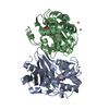
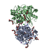
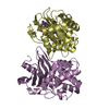
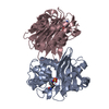
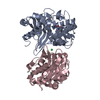
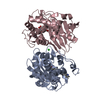
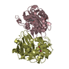
Journal: FEBS J / Year: 2018 Title: The biological assembly of OXA-48 reveals a dimer interface with high charge complementarity and very high affinity. Authors: Bjarte Aarmo Lund / Ane Molden Thomassen / Birgit Helene Berg Nesheim / Trine Josefine Olsen Carlsen / Johan Isaksson / Tony Christopeit / Hanna-Kirsti S Leiros / Abstract: Many class D β-lactamases form dimers in solution. The functional basis of the dimerization of OXA-48-like class D β-lactamases is not known, but in order to understand the structural requirements ...Many class D β-lactamases form dimers in solution. The functional basis of the dimerization of OXA-48-like class D β-lactamases is not known, but in order to understand the structural requirements for dimerization of OXA-48, we have characterized the dimer interface. Size exclusion chromatography, small angle X-ray scattering (SAXS), and nuclear magnetic resonance (NMR) were used to confirm the oligomeric state of OXA-48 in solution. X-ray crystallographic structures were used to elucidate the key interactions of dimerization. In silico residue scanning combined with site-directed mutagenesis was used to probe hot spots of dimerization. The affinity of dimerization was quantified using microscale thermophoresis, and the overall thermostability was investigated using differential scanning calorimetry. OXA-48 was consistently found to be a dimer in solution regardless of the method used, and the biological assembly found from the SAXS envelope is consistent with the dimer identified from the crystal structures. The buried chloride that interacts with Arg206 and Arg206' at the dimer interface was found to enhance the thermal stability by > 4 °C and crystal structures and mutations (R189A, R189A/R206A) identified several additional important ionic interactions. The affinity for OXA-48 R206A dimerization was in the picomolar range, thus revealing very high dimer affinity. In summary, OXA-48 has a very stable dimer interface, facilitated by noncovalent and predominantly charged interactions, which is stronger than the dimer interfaces previously described for other class D β-lactamases. PDB CODES: The oxacillinase-48 (OXA-48) R206A structure has PDB ID: 5OFT and OXA-48 R189A has PDB ID: 6GOA.
Contact author
Bjarte Aarmo Lund (UiT, UiT - The Arctic University of Norway, Tromso, Norway)
Title: Dimeric class D beta-lactamase OXA-48 / Measurement date: Feb 23, 2017 / Storage temperature: 25 °C / Cell temperature: 25 °C / Exposure time: 10 sec. / Number of frames: 10 / Unit: 1/nm /
Min
Max
Q
0.0788
4.8993
Distance distribution function P(R)
Sofotware P(R): GNOM 5.0 / Number of points: 671 /
Min
Max
Q
0.0835416
3.23758
P(R) point
1
671
R
0
7.35
Result
Type of curve: merged /
Experimental
Porod
MW
50 kDa
46 kDa
Volume
-
74 nm3
P(R)
Guinier
Guinier error
Forward scattering, I0
26.79
27.04
0.052
Radius of gyration, Rg
2.476 nm
2.47 nm
0.018
Min
Max
D
-
7.35
Guinier point
2
96
+
About Yorodumi
-
News
-
Feb 9, 2022. New format data for meta-information of EMDB entries
New format data for meta-information of EMDB entries
Version 3 of the EMDB header file is now the official format.
The previous official version 1.9 will be removed from the archive.
In the structure databanks used in Yorodumi, some data are registered as the other names, "COVID-19 virus" and "2019-nCoV". Here are the details of the virus and the list of structure data.
Jan 31, 2019. EMDB accession codes are about to change! (news from PDBe EMDB page)
EMDB accession codes are about to change! (news from PDBe EMDB page)
The allocation of 4 digits for EMDB accession codes will soon come to an end. Whilst these codes will remain in use, new EMDB accession codes will include an additional digit and will expand incrementally as the available range of codes is exhausted. The current 4-digit format prefixed with “EMD-” (i.e. EMD-XXXX) will advance to a 5-digit format (i.e. EMD-XXXXX), and so on. It is currently estimated that the 4-digit codes will be depleted around Spring 2019, at which point the 5-digit format will come into force.
The EM Navigator/Yorodumi systems omit the EMD- prefix.
Related info.:Q: What is EMD? / ID/Accession-code notation in Yorodumi/EM Navigator
Yorodumi is a browser for structure data from EMDB, PDB, SASBDB, etc.
This page is also the successor to EM Navigator detail page, and also detail information page/front-end page for Omokage search.
The word "yorodu" (or yorozu) is an old Japanese word meaning "ten thousand". "mi" (miru) is to see.
Related info.:EMDB / PDB / SASBDB / Comparison of 3 databanks / Yorodumi Search / Aug 31, 2016. New EM Navigator & Yorodumi / Yorodumi Papers / Jmol/JSmol / Function and homology information / Changes in new EM Navigator and Yorodumi
 Movie
Movie Controller

 Open data
 Basic information
 Sample
 Function and homology information
 Klebsiella pneumoniae (bacteria)
 Citation
 Contact author
 Structure visualization
 Downloads & links
 SASDEM3
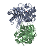


 Search similar-shape structures of this assembly by Omokage search (details)
 / Type of source: X-ray synchrotron / Wavelength: 0.09919 Å / Dist. spec. to detc.: 2.867 mm


