+Search query
-Structure paper
| Title | The biological assembly of OXA-48 reveals a dimer interface with high charge complementarity and very high affinity. |
|---|---|
| Journal, issue, pages | FEBS J, Vol. 285, Issue 22, Page 4214-4228, Year 2018 |
| Publish date | Sep 7, 2018 |
 Authors Authors | Bjarte Aarmo Lund / Ane Molden Thomassen / Birgit Helene Berg Nesheim / Trine Josefine Olsen Carlsen / Johan Isaksson / Tony Christopeit / Hanna-Kirsti S Leiros /  |
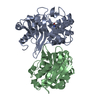
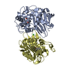
| PubMed Abstract | Many class D β-lactamases form dimers in solution. The functional basis of the dimerization of OXA-48-like class D β-lactamases is not known, but in order to understand the structural requirements ...Many class D β-lactamases form dimers in solution. The functional basis of the dimerization of OXA-48-like class D β-lactamases is not known, but in order to understand the structural requirements for dimerization of OXA-48, we have characterized the dimer interface. Size exclusion chromatography, small angle X-ray scattering (SAXS), and nuclear magnetic resonance (NMR) were used to confirm the oligomeric state of OXA-48 in solution. X-ray crystallographic structures were used to elucidate the key interactions of dimerization. In silico residue scanning combined with site-directed mutagenesis was used to probe hot spots of dimerization. The affinity of dimerization was quantified using microscale thermophoresis, and the overall thermostability was investigated using differential scanning calorimetry. OXA-48 was consistently found to be a dimer in solution regardless of the method used, and the biological assembly found from the SAXS envelope is consistent with the dimer identified from the crystal structures. The buried chloride that interacts with Arg206 and Arg206' at the dimer interface was found to enhance the thermal stability by > 4 °C and crystal structures and mutations (R189A, R189A/R206A) identified several additional important ionic interactions. The affinity for OXA-48 R206A dimerization was in the picomolar range, thus revealing very high dimer affinity. In summary, OXA-48 has a very stable dimer interface, facilitated by noncovalent and predominantly charged interactions, which is stronger than the dimer interfaces previously described for other class D β-lactamases. PDB CODES: The oxacillinase-48 (OXA-48) R206A structure has PDB ID: 5OFT and OXA-48 R189A has PDB ID: 6GOA. |
 External links External links |  FEBS J / FEBS J /  PubMed:30153368 PubMed:30153368 |
| Methods | SAS (X-ray synchrotron) / X-ray diffraction |
| Resolution | 2.55 - 3.2 Å |
| Structure data |  SASDEM3:  PDB-5oft:  PDB-6goa: |
| Chemicals |  ChemComp-HOH:  ChemComp-CL: |
| Source |
|
 Keywords Keywords | ANTIBIOTIC / dimerization / OXA-48 / Oxacillinase / carbapenemase / beta-lactamase / HYDROLASE |
 Movie
Movie Controller
 Structure viewers
 About Yorodumi Papers


 klebsiella pneumoniae (bacteria)