 Movie
Movie Controller

+ Open data

- Basic information
| Entry | Database: SASBDB / ID: SASDDE6 |
|---|---|
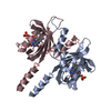
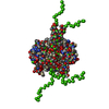
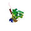
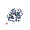
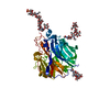
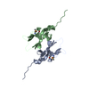
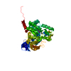
 Sample Sample | Photoreceptor sensory box 1 light-state (SB1-LOV-R66I)
|
| Function / homology |  Function and homology information Function and homology information |
| Biological species |  Pseudomonas putida (strain ATCC 47054 / DSM 6125 / NCIMB 11950 / KT2440) (bacteria) Pseudomonas putida (strain ATCC 47054 / DSM 6125 / NCIMB 11950 / KT2440) (bacteria) |
 Citation Citation | Journal: PLoS One / Year: 2018 Title: Small-angle X-ray scattering study of the kinetics of light-dark transition in a LOV protein. Authors: Katrin Röllen / Joachim Granzin / Renu Batra-Safferling / Andreas Maximilian Stadler /  Abstract: Light, oxygen, voltage (LOV) photoreceptors consist of conserved photo-responsive domains in bacteria, archaea, plants and fungi, and detect blue-light via a flavin cofactor. We investigated the blue- ...Light, oxygen, voltage (LOV) photoreceptors consist of conserved photo-responsive domains in bacteria, archaea, plants and fungi, and detect blue-light via a flavin cofactor. We investigated the blue-light induced conformational transition of the dimeric photoreceptor PpSB1-LOV-R66I from Pseudomonas putida in solution by using small-angle X-ray scattering (SAXS). SAXS experiments of the fully populated light- and dark-states under steady-state conditions revealed significant structural differences between the two states that are in agreement with the known structures determined by crystallography. We followed the transition from the light- to the dark-state by using SAXS measurements in real-time. A two-state model based on the light- and dark-state conformations could describe the measured time-course SAXS data with a relaxation time τREC of ~ 34 to 35 min being larger than the recovery time found with UV/vis spectroscopy. Unlike the flavin chromophore-based UV/vis method that is sensitive to the local chromophore environment in flavoproteins, SAXS-based assay depends on protein conformational changes and provides with an alternative to measure the recovery kinetics. |
 Contact author Contact author |
|
- Structure visualization
| Structure viewer | Molecule: MolmilJmol/JSmol |
|---|
- Downloads & links
-Data source
| SASBDB page |  SASDDE6 SASDDE6 |
|---|
-Related structure data


-External links
| Related items in Molecule of the Month |
|---|


-Models
| Model #1970 |  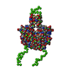 Type: mix / Radius of dummy atoms: 1.90 A / Chi-square value: 1.891  Search similar-shape structures of this assembly by Omokage search (details) Search similar-shape structures of this assembly by Omokage search (details) |
|---|---|
| Model #1971 |   Type: mix / Software: (2.8.3) / Radius of dummy atoms: 1.90 A / Chi-square value: 5.639 Search similar-shape structures of this assembly by Omokage search (details) |
| Model #1972 | 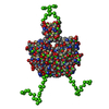 Type: mix / Radius of dummy atoms: 1.90 A / Chi-square value: 5.639 Search similar-shape structures of this assembly by Omokage search (details) |
| Model #1973 |  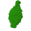 Type: dummy / Software: (2.8.3) / Radius of dummy atoms: 2.00 A / Symmetry: P2 / Chi-square value: 0.783 Search similar-shape structures of this assembly by Omokage search (details) |
-Sample
| Sample | Name: Photoreceptor sensory box 1 light-state (SB1-LOV-R66I) Specimen concentration: 1.05-6.10 |
|---|---|
| Buffer | Name: 10mM Tris, 10 mM NaCl / pH: 7 |
| Entity #1060 | Name: PpSB1-LOV-R66I light / Type: protein / Description: Sensory box protein light-state (R66I) / Formula weight: 18.588 / Num. of mol.: 2 Source: Pseudomonas putida (strain ATCC 47054 / DSM 6125 / NCIMB 11950 / KT2440) References: UniProt: Q88E39 Sequence: MGSSHHHHHH SSGLVPRGSH MINAQLLQSM VDASNDGIVV AEKEGDDTIL IYVNAAFEYL TGYSRDEILY QDCRFLQGDD RDQLGRARIR KAMAEGRPCR EVLRNYRKDG SAFWNELSIT PVKSDFDQRT YFIGIQKDVS RQVELERELA ELRARPKPDE RA |
-Experimental information
| Beam | Instrument name: ESRF BM29 / City: Grenoble / 国: France  / Type of source: X-ray synchrotron / Wavelength: 0.099 Å / Dist. spec. to detc.: 2.867 mm / Type of source: X-ray synchrotron / Wavelength: 0.099 Å / Dist. spec. to detc.: 2.867 mm | ||||||||||||||||||||||||||||||||||||
|---|---|---|---|---|---|---|---|---|---|---|---|---|---|---|---|---|---|---|---|---|---|---|---|---|---|---|---|---|---|---|---|---|---|---|---|---|---|
| Detector | Name: Pilatus 1M / Pixsize x: 172 mm | ||||||||||||||||||||||||||||||||||||
| Scan |  Measurement date: Dec 2, 2014 / Storage temperature: 10 °C / Cell temperature: 10 °C / Exposure time: 1 sec. / Number of frames: 10 / Unit: 1/nm /
| ||||||||||||||||||||||||||||||||||||
| Distance distribution function P(R) | 
| ||||||||||||||||||||||||||||||||||||
| Result | 
|
