 ムービー
ムービー コントローラー

+ データを開く

- 基本情報
基本情報
| 登録情報 | データベース: SASBDB / ID: SASDDD9 |
|---|---|
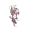
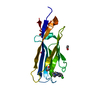
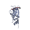
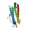
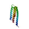
 試料 試料 | Cardiac myosin binding protein C: tri-helix bundle from the motif with C2 domain
|
| 機能・相同性 |  機能・相同性情報 機能・相同性情報C zone / regulation of muscle filament sliding / striated muscle myosin thick filament / A band / regulation of striated muscle contraction / cardiac myofibril / Striated Muscle Contraction / M band / regulation of cardiac muscle cell contraction / structural constituent of muscle ...C zone / regulation of muscle filament sliding / striated muscle myosin thick filament / A band / regulation of striated muscle contraction / cardiac myofibril / Striated Muscle Contraction / M band / regulation of cardiac muscle cell contraction / structural constituent of muscle / sarcomere organization / myosin heavy chain binding / ventricular cardiac muscle tissue morphogenesis / myosin binding / ATPase activator activity / heart morphogenesis / cardiac muscle contraction / titin binding / sarcomere / actin binding / cell adhesion / metal ion binding / identical protein binding / cytosol 類似検索 - 分子機能 |
| 生物種 | human sequence obtained using E. coli expression |
 引用 引用 | ジャーナル: Structure / 年: 2016 タイトル: A Highly Conserved Yet Flexible Linker Is Part of a Polymorphic Protein-Binding Domain in Myosin-Binding Protein C. 著者: Katharine A Michie / Ann H Kwan / Chang-Shung Tung / J Mitchell Guss / Jill Trewhella /   要旨: The nuclear magnetic resonance (NMR) structure of the tri-helix bundle (THB) of the m-domain plus C2 (ΔmC2) of myosin-binding protein C (MyBP-C) has revealed a highly flexible seven-residue linker ...The nuclear magnetic resonance (NMR) structure of the tri-helix bundle (THB) of the m-domain plus C2 (ΔmC2) of myosin-binding protein C (MyBP-C) has revealed a highly flexible seven-residue linker between the structured THB and C2. Bioinformatics shows significant patterns of conservation across the THB-linker sequence, with the linker containing a strictly conserved serine in all MyBP-C isoforms. Clinically linked mutations further support the functional significance of the THB-linker region. NMR, small-angle X-ray scattering, and binding studies show the THB-linker plus the first ten residues of C2 undergo dramatic changes when ΔmC2 binds Ca-calmodulin, with the linker and C2 N-terminal residues contributing significantly to the affinity. Modeling of all available experimental data indicates that the THB tertiary structure must be disrupted to form the complex. These results are discussed in the context of the THB-linker and the N-terminal residues of C2 forming a polymorphic binding domain that could accommodate multiple binding partners in the dynamic sarcomere. |
 登録者 登録者 |
|
- 構造の表示
構造の表示
| 構造ビューア | 分子: MolmilJmol/JSmol |
|---|
- ダウンロードとリンク
 SASDDD9


-モデル
| モデル #2155 |   タイプ: atomic コメント: one state of 2-state MultiFoXS model, weight 0.753 カイ2乗値: 3.56671154143 / P-value: 0.820000  Omokage検索でこの集合体の類似形状データを探す (詳細) Omokage検索でこの集合体の類似形状データを探す (詳細) |
|---|---|
| モデル #2156 |  タイプ: atomic コメント: one state of 2-state MultiFoXS model, weight 0.247 カイ2乗値: 3.56671154143 / P-value: 0.820000 Omokage検索でこの集合体の類似形状データを探す (詳細) |
-試料
| 試料 | 名称: Cardiac myosin binding protein C: tri-helix bundle from the motif with C2 domain 試料濃度: 0.98-4.89 |
|---|---|
| バッファ | 名称: 150 mM NaCl, 10 mM MES, 2 mM TCEP, 1 mM NaN3 at 4°C pH: 6.5 |
| 要素 #1178 | 名称: tri-helix bundle-C2 / タイプ: protein 記述: cardiac myosin binding protein C: tri-helix bundle-C2 分子量: 15.474 / 分子数: 1 / 由来: human sequence obtained using E. coli expression / 参照: UniProt: Q14896 配列: GPGSEDVWEI LRQAPPSEYE RIAFQYGVTD LRGMLKRLKG MRRDEKKSTA FQKKLEPAYQ VSKGHKIRLT VELADHDAEV KWLKNGQEIQ MSGSKYIFES IGAKRTLTIS QCSLADDAAY QCVVGGEKCS TELFVKE |
-実験情報
| ビーム | 設備名称: Australian Synchrotron SAXS/WAXS / 地域: Melbourne / 国: Australia / 形状: Point / 線源: X-ray synchrotron / 波長: 0.10332 Å / スペクトロメータ・検出器間距離: 2.68024 mm | ||||||||||||||||||||||||||||||||||||
|---|---|---|---|---|---|---|---|---|---|---|---|---|---|---|---|---|---|---|---|---|---|---|---|---|---|---|---|---|---|---|---|---|---|---|---|---|---|
| 検出器 | 名称: Pilatus 1M / Pixsize x: 172 mm | ||||||||||||||||||||||||||||||||||||
| スキャン |  測定日: 2015年4月18日 / セル温度: 22 °C / 照射時間: 1 sec. / フレーム数: 26 / 単位: 1/A /
| ||||||||||||||||||||||||||||||||||||
| 距離分布関数 P(R) | 
| ||||||||||||||||||||||||||||||||||||
| 結果 |  コメント: The concentration series SAXS data and EOM models are provided in the full entry zip archive.
|
