[English] 日本語
 Yorodumi
- SASDAW5: XMRV RT + DNA/RNA hybrid (apo XMRV RT, XM + RNA_DNA hybrid substr... -
+ Open data
- Basic information
| Entry | Database: SASBDB / ID: SASDAW5 |
|---|---|
 Sample Sample | XMRV RT + DNA/RNA hybrid
|
| Biological species |  |
 Citation Citation |  Journal: Nucleic Acids Res / Year: 2013 Journal: Nucleic Acids Res / Year: 2013Title: Structural analysis of monomeric retroviral reverse transcriptase in complex with an RNA/DNA hybrid. Authors: Elzbieta Nowak / Wojciech Potrzebowski / Petr V Konarev / Jason W Rausch / Marion K Bona / Dmitri I Svergun / Janusz M Bujnicki / Stuart F J Le Grice / Marcin Nowotny /  Abstract: A key step in proliferation of retroviruses is the conversion of their RNA genome to double-stranded DNA, a process catalysed by multifunctional reverse transcriptases (RTs). Dimeric and monomeric ...A key step in proliferation of retroviruses is the conversion of their RNA genome to double-stranded DNA, a process catalysed by multifunctional reverse transcriptases (RTs). Dimeric and monomeric RTs have been described, the latter exemplified by the enzyme of Moloney murine leukaemia virus. However, structural information is lacking that describes the substrate binding mechanism for a monomeric RT. We report here the first crystal structure of a complex between an RNA/DNA hybrid substrate and polymerase-connection fragment of the single-subunit RT from xenotropic murine leukaemia virus-related virus, a close relative of Moloney murine leukaemia virus. A comparison with p66/p51 human immunodeficiency virus-1 RT shows that substrate binding around the polymerase active site is conserved but differs in the thumb and connection subdomains. Small-angle X-ray scattering was used to model full-length xenotropic murine leukaemia virus-related virus RT, demonstrating that its mobile RNase H domain becomes ordered in the presence of a substrate-a key difference between monomeric and dimeric RTs. |
 Contact author Contact author |
|
- Structure visualization
| Structure viewer | Molecule:  Molmil Molmil Jmol/JSmol Jmol/JSmol |
|---|
- Downloads & links
-Models
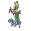
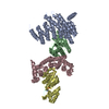
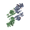
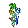
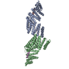
| Model #142 |  Type: atomic / Software: CRYSOL / Chi-square value: 0.855625  Search similar-shape structures of this assembly by Omokage search (details) Search similar-shape structures of this assembly by Omokage search (details) |
|---|
- Sample
 Sample Sample | Name: XMRV RT + DNA/RNA hybrid / Sample MW: 90 kDa / Entity id: 103 / 112 |
|---|---|
| Buffer | Name: 10mM HEPES / pH: 6.5 / Composition: KCl 100.000 mM, glycerol 5.000 % |
| Entity #103 | Name: XM / Type: protein / Description: apo XMRV RT / Formula weight: 75 / Num. of mol.: 1 / Source: Escherichia coli Sequence: MSHHHHHHSA TLNIEDEYRL HETSKEPDVP LGSTWLSDFP QAWAETGGMG LAVRQAPLII PLKATSTPVS IKQYPMSQEA RLGIKPHIQR LLDQGILVPC QSPWNTPLLP VKKPGTNDYR PVQDLREVNK RVEDIHPTVP NPYNLLSGLP PSHQWYTVLD LKDAFFCLRL ...Sequence: MSHHHHHHSA TLNIEDEYRL HETSKEPDVP LGSTWLSDFP QAWAETGGMG LAVRQAPLII PLKATSTPVS IKQYPMSQEA RLGIKPHIQR LLDQGILVPC QSPWNTPLLP VKKPGTNDYR PVQDLREVNK RVEDIHPTVP NPYNLLSGLP PSHQWYTVLD LKDAFFCLRL HPTSQPLFAF EWRDPEMGIS GQLTWTRLPQ GFKNSPTLFD EALHRDLADF RIQHPDLILL QYVDDLLLAA TSEQDCQRGT RALLQTLGNL GYRASAKKAQ ICQKQVKYLG YLLKEGQRWL TEARKETVMG QPTPKTPRQL REFLGTAGFC RLWIPGFAEM AAPLYPLTKT GTLFNWGPDQ QKAYQEIKQA LLTAPALGLP DLTKPFELFV DEKQGYAKGV LTQKLGPWRR PVAYLSKKLD PVAAGWPPCL RMVAAIAVLT KDAGKLTMGQ PLVILAPHAV EALVKQPPDR WLSNARMTHY QAMLLDTDRV QFGPVVALNP ATLLPLPEKE APHDCLEILA ETHGTRPDLT DQPIPDADYT WYTDGSSFLQ EGQRRAGAAV TTETEVIWAR ALPAGTSAQR AELIALTQAL KMAEGKKLNV YTDSRYAFAT AHVHGEIYRR RGLLTSEGRE IKNKNEILAL LKALFLPKRL SIIHCPGHQK GNSAEARGNR MADQAAREAA MKAVLETSTL L |
| Entity #112 | Name: PPT18 / Type: other / Description: RNA_DNA hybrid substrate / Formula weight: 15 / Num. of mol.: 1 Sequence: 5UAGUCUCCA GAAAAAGGGG GGAAUG35AT TCCCCCCTTT TTCTGGAGAC 3 |
-Experimental information
| Beam | Instrument name:  DORIS III X33 DORIS III X33  / City: Hamburg / 国: Germany / City: Hamburg / 国: Germany  / Shape: 0.6 / Type of source: X-ray synchrotron / Wavelength: 0.15 Å / Dist. spec. to detc.: 2.7 mm / Shape: 0.6 / Type of source: X-ray synchrotron / Wavelength: 0.15 Å / Dist. spec. to detc.: 2.7 mm | |||||||||||||||||||||||||||
|---|---|---|---|---|---|---|---|---|---|---|---|---|---|---|---|---|---|---|---|---|---|---|---|---|---|---|---|---|
| Detector | Name: Pilatus 1M-W / Pixsize x: 0.172 mm | |||||||||||||||||||||||||||
| Scan |
| |||||||||||||||||||||||||||
| Distance distribution function P(R) |
| |||||||||||||||||||||||||||
| Result |
|
 Movie
Movie Controller

 SASDAW5


